
Figure 4.
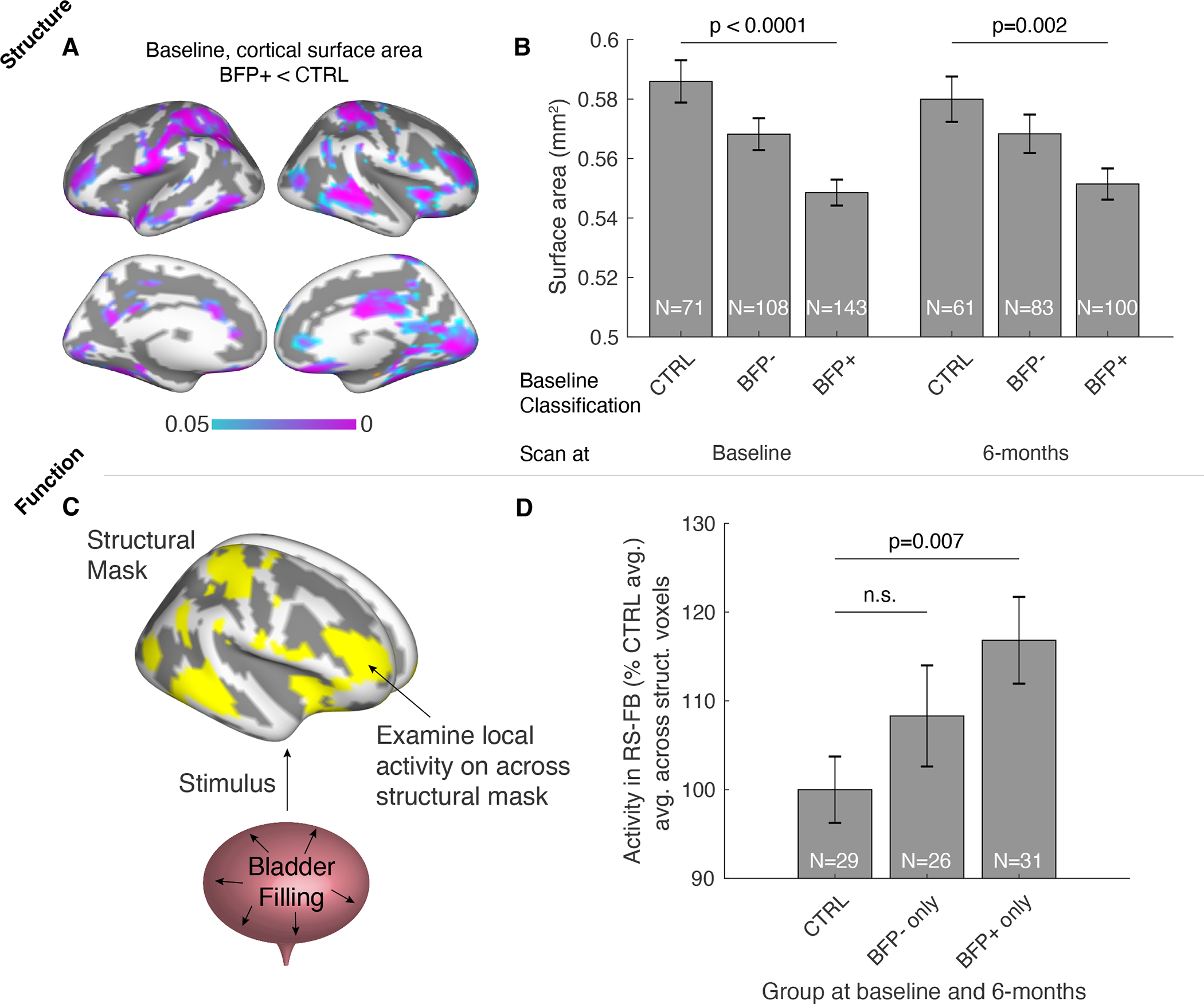
Changes in brain morphology and bladder filling function in BFP+ UCPPS patients. A. Cortical surface area decreases were observed at baseline in BFP+ patients compared to controls (p<0.05, FDR corrected for multiple comparisons across cortical mesh vertices). B. Decreases observed at baseline were observed again in the same individuals imaged 6-months later. C. Functional activity changes were observed across the brain masked to regions expressing structural changes shown in A. D. Activity was significantly higher during resting-state with full bladder (RS-FB) in BFP+ patients compared to controls (meanFD<0.20 shown here, see Supplemental Figure 1).