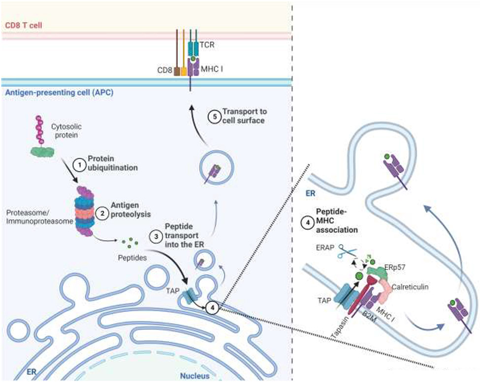
Figure-1:
The MHC I antigen presentation pathway. Cytosolic and nuclear proteins are degraded by proteasomes and immunoproteasomes into oligopeptides. Some of these peptides are then translocated into the endoplasmic reticulum (ER) by the TAP transporter. In the ER, ERAPs may further trim these oligopeptides, and then ones of the right length and sequence bind to MHC I molecules within a peptide-loading complex, which contains Tapasin, TAP, calreticulin, and ERP57. Peptide-loaded MHCI molecules are then transported to the cell surface for display to CD8+ T cells. Created with BioRender.com.

