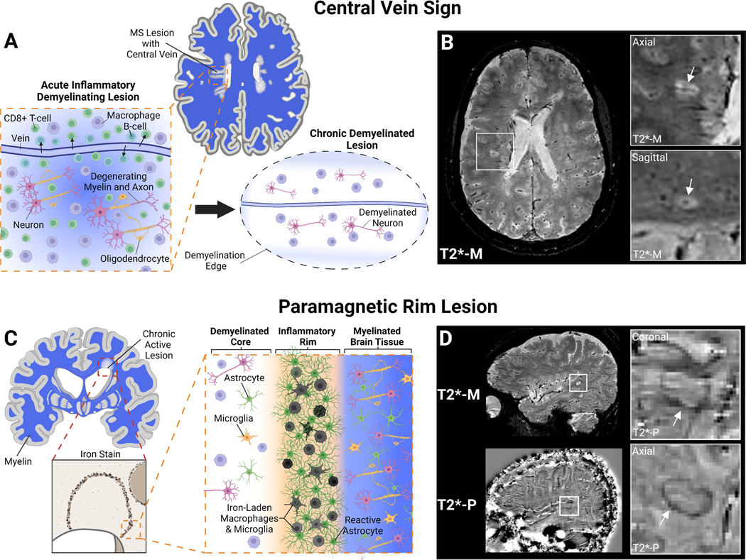
Figure 1: Emerging MRI Biomarkers in MS: Central Vein Sign and Paramagnetic Rim Lesions.
A) White matter lesions in MS are acutely result of immune cell infiltration, particularly CD8+ T-cells, from the periphery into the CNS via small penetrating veins. These inflammatory lesions result in oligodendrocyte and myelin damage as well as neuro-axonal degeneration. After peripheral lymphocyte infiltration resolves a chronic demyelinated lesion centered around a vein remains B) Certain MRI sequences can depict white matter pathology and small CNS vessels simultaneously (e.g., T2*-weighted magnitude reconstruction; T2*-M). These small veins within classic ovoid MS lesions can be visualized and quantified to aid in MS diagnosis. Inserts show confirmation of central vein in two planes. C) Chronic active lesions in MS can be identified pathologically by an iron-rim at the lesion edge that contains iron-laden macrophages and microglia as well as activated astrocytes. D) These iron-rimed chronic active lesions can be visualized on MRI as paramagnetic rim lesions (PRLs) by “unwrapping” the phase reconstruction of the same T2*-weighted imaging (T2*-P)175. Paramagnetic rim lesions may represent a biomarker of at least one cause of progressive disease in MS. Created with BioRender.com

