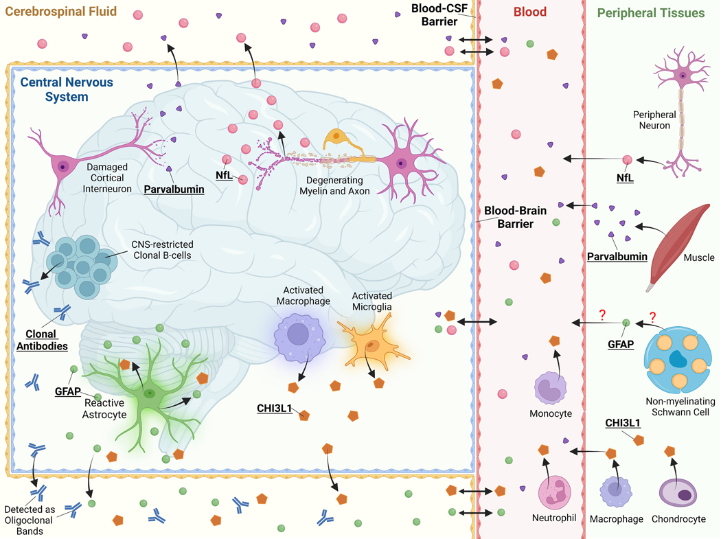
Figure 2: Emerging Neuroglial Biomarkers in MS.
Schematic of the CNS, periphery, and blood-brain barrier, and blood-CSF barrier cell types relevant to emerging neuroglial biomarkers and CSF-specific oligoclonal bands. Released neuroglial protein biomarkers are released from one or a select few CNS resident cell types where they can traffic to the CSF and blood. These cell-specific biomarkers may thus reflect cell-type specific pathology, such as axonal damage in the case of Nfl. Many neuroglial biomarkers also have identified or potential peripheral sources that may, if a significant source, limit or prevent the use of blood levels to be a useful as a biomarker, such as the case with parvalbumin and CHI3L1. Many neuroglial biomarkers cross the CSF-blood barrier and even the blood-brain barrier, particularly in the setting of blood-brain barrier injury such as occurs in an active MS lesion. This equilibrium between CSF and serum or plasma levels is important to determine for each biomarker as high peripheral levels from a non-CNS source may impact CSF levels requiring correction of obtained CSF values. Abbreviations: CHI3L1, chitinase-3 like protein 1; GFAP, glial fibrillary acidic protein; Nfl, neurofilament. Created with BioRender.com

