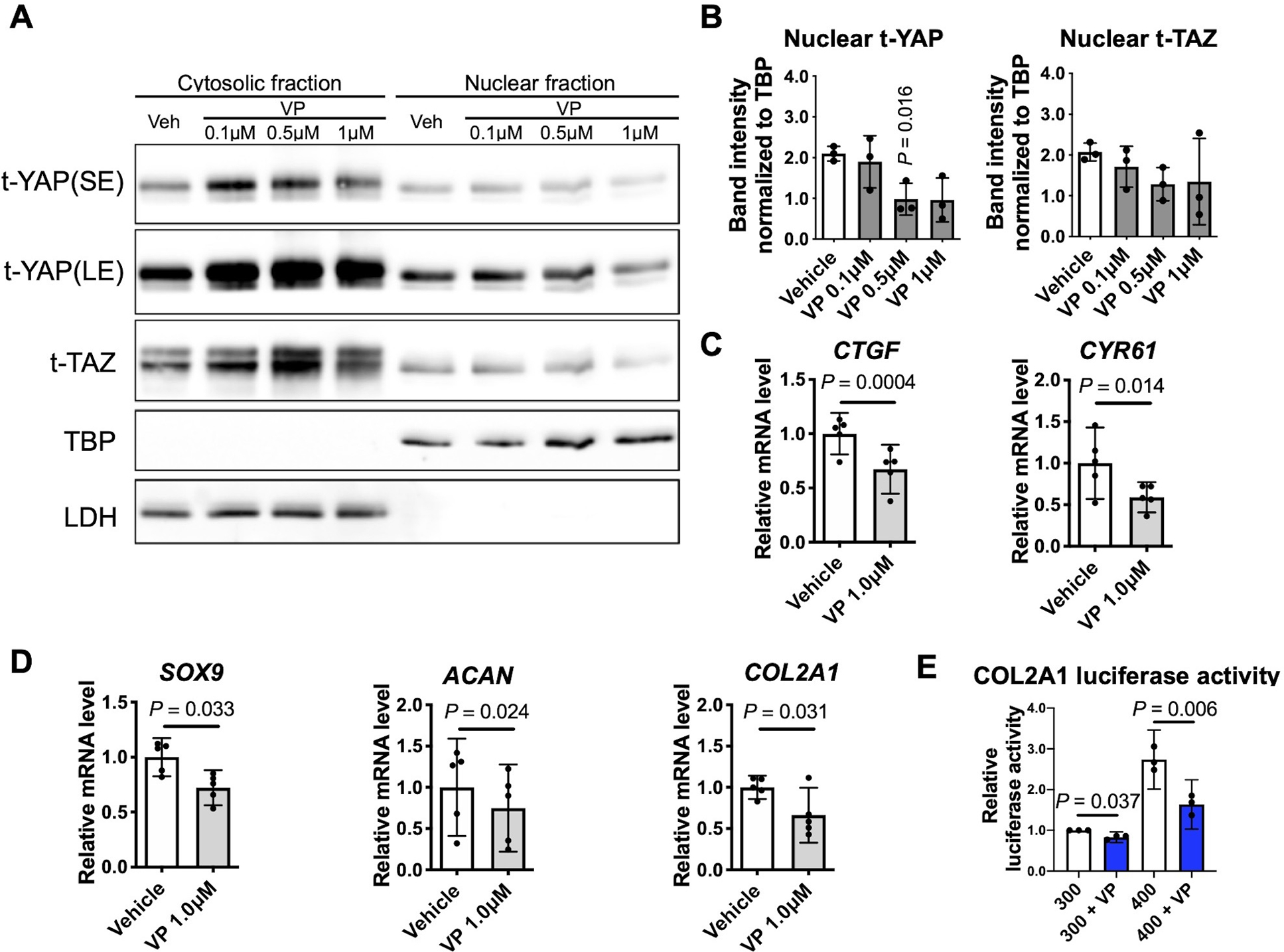
Fig. 3. Effects of YAP inhibition on chondrocyte anabolic activity.

(A) Chondrocytes incubated in standard 300 mOsm media were treated for 6 hr with the indicated concentration of the YAP inhibitor verteporfin (VP). Protein levels of YAP and TAZ in cytosolic and nuclear fractions were determined by immunoblotting. The immunoblots shown are representative of three independent donors (t, total; SE, short exposure; LE, long exposure). (B) Densitometry analysis of YAP/TAZ nuclear abundance. Band intensities of nuclear YAP (n=3 independent donors) and TAZ (n=3 independent donors) were normalized to the loading control TBP and are presented as the mean ± standard deviation; indicated p-values were determined by paired t-tests used to compare different conditions to vehicle controls. (C, D) Quantification of the expression of the indicated genes in response to 6 hr of 1 μM VP treatment. Data are presented as the mean with 95% confidence interval (CI). Differences from vehicle controls are indicated (n = 5 independent donors). (E) Chondrocytes were transiently transfected with the SOX9-dependent COL2A1 luciferase reporter construct for 48 hr, followed by incubation in 300 mOsm media or 400 mOsm media produced using sucrose for 12 h after pretreatment with VP at 1 μM for 6 h and measurement of luciferase activity. The data are expressed as fold changes relative to 300 mOsm controls (n = 3 independent donors). For each condition, one-sample t-test was applied to compare logged 2 transformed fold change to zero, which corresponds to the fold change of 1 indicating no difference.
