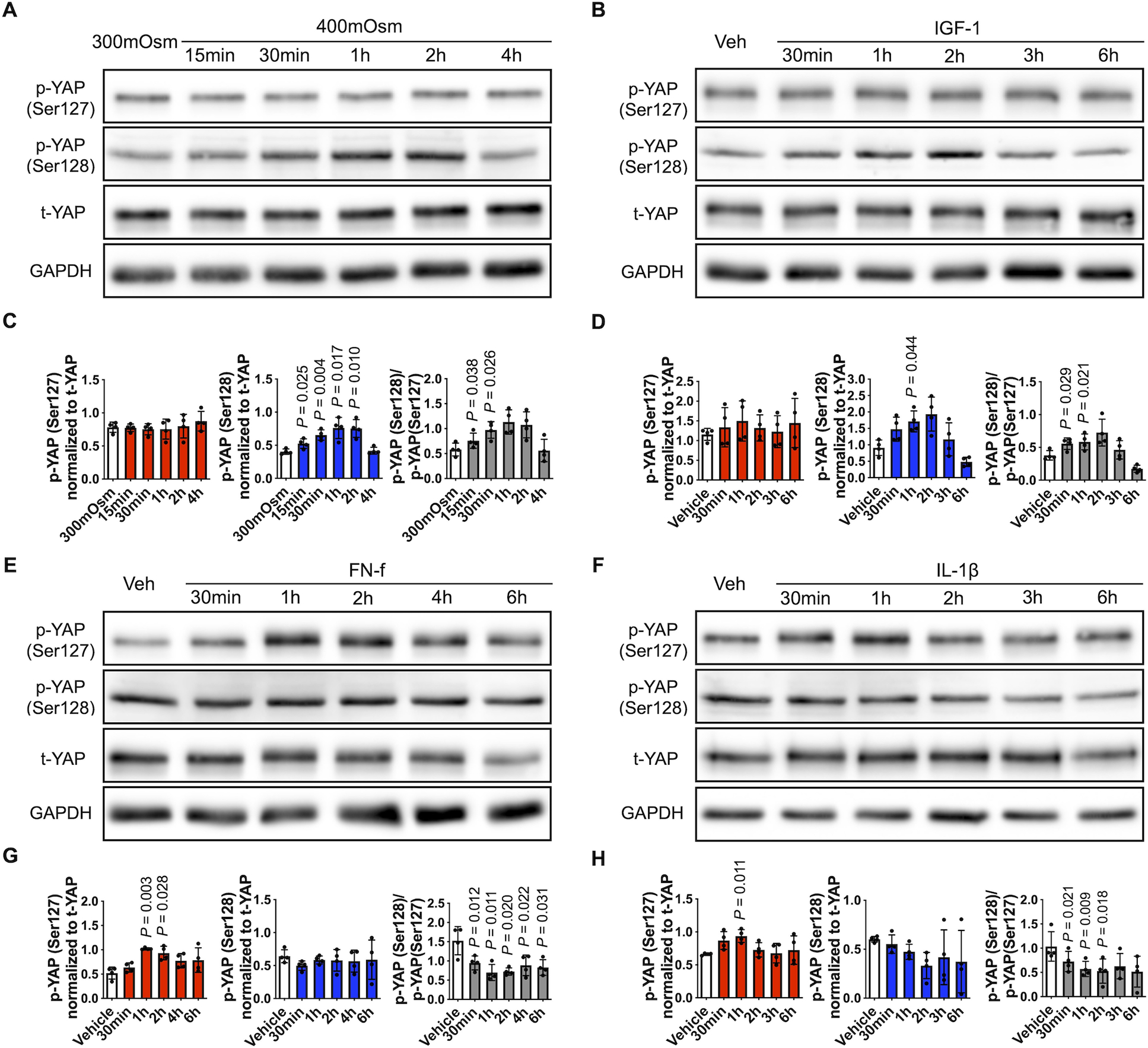
Fig. 6. YAP phosphorylation in response to changes in osmolarity, IGF-1, FN-f, and IL-1β.

(A) Chondrocytes were incubated in 300 mOsm media or 400 mOsm media generated using sucrose for the indicated times. (B, E, F) Chondrocytes incubated in 300 mOsm media were treated with IGF-1 (100 ng/ml), FN-f (1 μM) or IL-1β (10 ng/ml) for the indicated times. Cell lysates were prepared and subjected to immunoblot analysis for p-YAP (Ser127), p-YAP (Ser128) and t-YAP. The immunoblots shown are representative of four independent donors (p, phospho-; t, total). (C, D, G, H) Densitometric analysis showing YAP phosphorylation at Ser127 (red column) and Ser128 (blue column). Phosphorylated bands were normalized to total protein as loading controls. The ratio of p-YAP (Ser128) to p-YAP (Ser127) was also analyzed (gray column). Data are presented as the mean ± standard deviation; indicated p-values were determined by paired t-tests used to compare different conditions either to 300 mOsm controls or vehicle controls.
