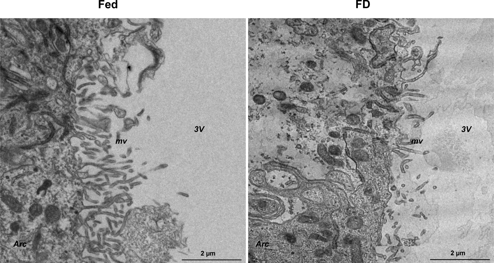
Figure 1:

Food deprivation reduces microvilli surface area of α2 tanycytes, suggesting decreased CSF–hypothalamus permeability. Transmission electron microscopy of α2 tanycytes in the mouse mediobasal hypothalamus bordering the 3V, ventromedial nucleus, and arcuate nucleus of the hypothalamus showed that, compared to control mice (Fed), mice that have been food deprived (FD) have shorter microvilli with markedly lower surface area facing the 3V. These data suggest that in the FD mice the diffusion of circulating factors is more restricted through the tanycytes that ultimately project to the Arc and 3V CSF. 3V, third ventricle; Arc, arcuate nucleus; FD, food deprivation; mv, microvilli; VMH, ventromedial nucleus. Scale bar = 2 μm.
