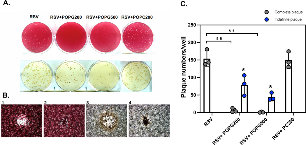
Figure 8. POPG inhibits RSV spread in HEp2 cells after infection was established.
HEp2 cells were challenged with RSV at a multiplicity of infection (m.o.i) of 10−4 for 2 h. Subsequently, the cells were washed and next overlayed with 0.3% agarose prepared in tissue culture medium either with, or without, or 200, or 500 μg/mL phospholipid (RSV, RSV+POPG200, RSV+POPG500, RSV+POPC200). Plaque numbers were quantified by staining with neutral red at day 6. Plaques in each condition were visualized with neutral red staining (panel A upper row) or were detected by immunostaining with antibody for RSV F protein11 (panel A lower row). Panel B shows images under microscopy with neutral red (panel B-1 and B-2), or with anti-RSV antibody (panel B-3 and B-4) 11. The quantitative data for plaque numbers are shown in Fig. 8C and the data are shown as mean ± SD, §§ indicates p<0.001 by paired t-test.

