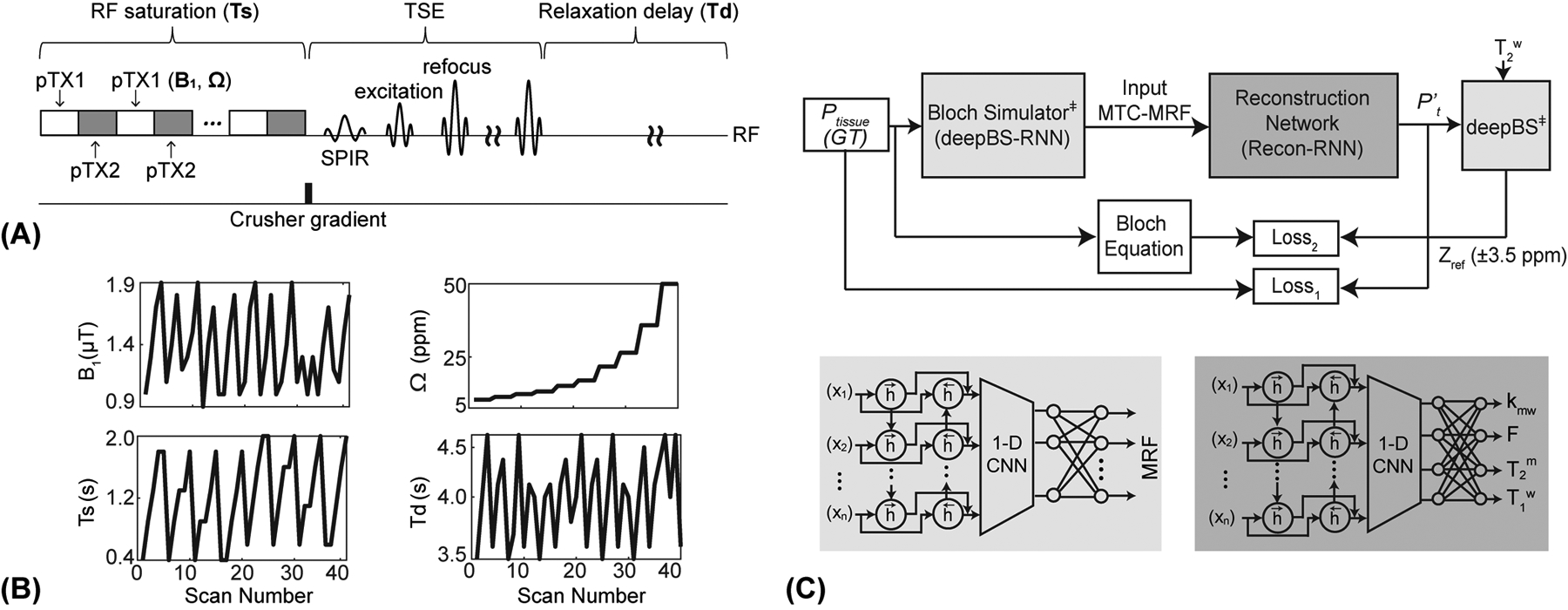
Figure 1.

(A) An illustration of RF saturation-encoded MTC-MRF sequence. A two-channel parallel transmission (pTX) was used to achieve continuous RF saturation at a 100% duty cycle. A variable density under-sampling pattern with the elliptic-centric k-space ordering was used in a turbo spin-echo (TSE) sequence and a spectral pre-saturation with inversion recovery (SPIR) was used for fat-suppressed data acquisitions. (B) An example of B1, Ts, Ω, and Td schedule for an MRF image acquisition. (C) MTC-MRF framework consisting of a deep Bloch simulator (deepBS-RNN) and a reconstruction network (Recon-RNN). Pt represents ground-truth (GT) tissue parameters and P’t represents estimated tissue parameters from the Recon-RNN. The estimated tissue parameters (P’t) and acquired T2w are fed to the pre-trained deep Bloch simulator (deepBS) module as input to calculate baseline reference signals (Zref(±3.5 ppm) = 100% - MTC (±3.5 ppm)). ‡ indicates pre-trained neural networks.
