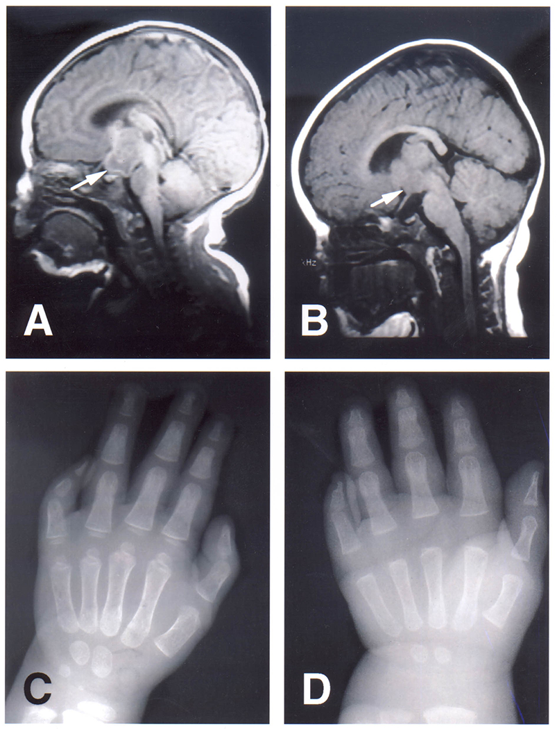
Figure 2:

Sagittal T1-weighted MRI of the brain in individual 1 (A) at the age of 2 weeks and in individual 2 (B) at the age of 7 months. Note the interpeduncular mass in continuity with the hypothalamus (arrows). The masses are isointense to grey matter. X-rays of the left hands of individual 1 (C) at the age of 2 years and of individual 2 (D) at the age of 7 months. Note the pronounced shortness of especially the middle and distal phalanx of the fifth finger. The middle and distal phalanx in individual 2 appear to show symphalangism.
