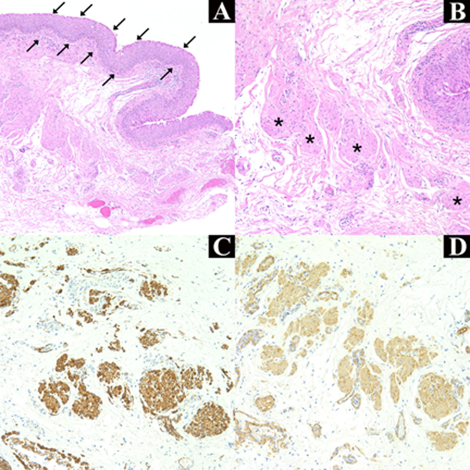
Figure 2. Histopathology of Conjunctival Lesion.

Acanthotic mucosal epithelium with squamous metaplasia (arrows) with underlying fibrovascular tissue within the substantia propria is present (A) (H&E, original magnification, × 100). Higher power demonstrates loose clusters of poorly circumscribed cells with eosinophilic cytoplasm and spindle-shaped nuclei consistent with smooth muscle (asterisks) (B) (H&E, original magnification × 200). Desmin (C) and smooth muscle actin (D) immunohistochemical stains are positive (original magnification, × 200).
