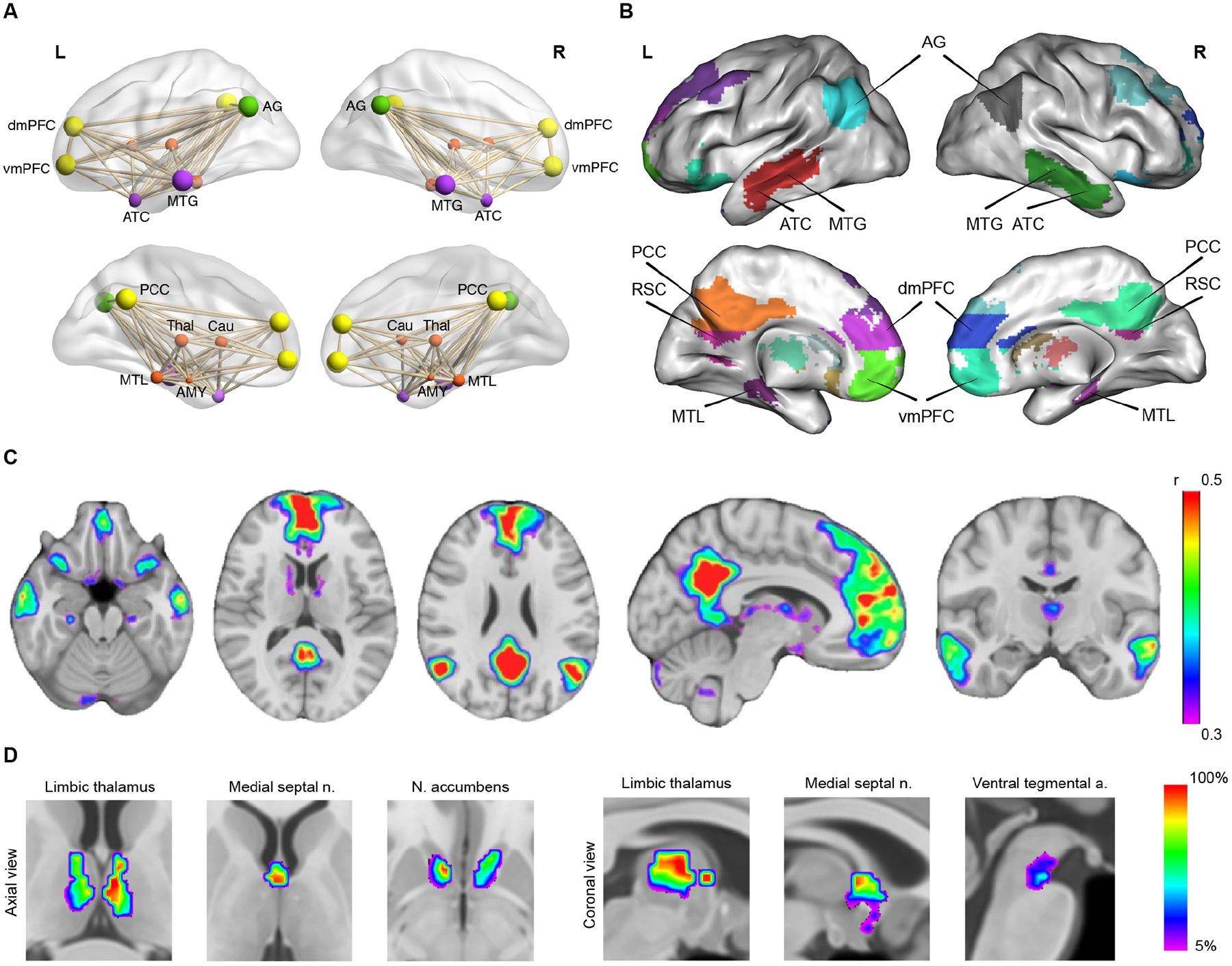
Figure 1. Cortical and subcortical nodes of the DMN.

(A) Illustration of DMN nodes as a functionally and anatomically interconnected system. (B, C) Cortical nodes of the DMN: posterior cingulate cortex (PCC) and retrosplenial cortex (RSC) in posterior medial parietal cortex; medial PFC (mPFC) with its dorsomedial (dmPFC) and ventromedial (dmPFC) subdivisions; anterior temporal cortex (ATC); middle temporal gyrus (MTG) in lateral temporal cortex; medial temporal lobe (MTL); and angular gyrus (AG) in lateral parietal cortex. (D) Subcortical nodes of the DMN: anterior and mediodorsal thalamic nuclei, medial septal nuclei, and nucleus accumbens. Adapted from3.
