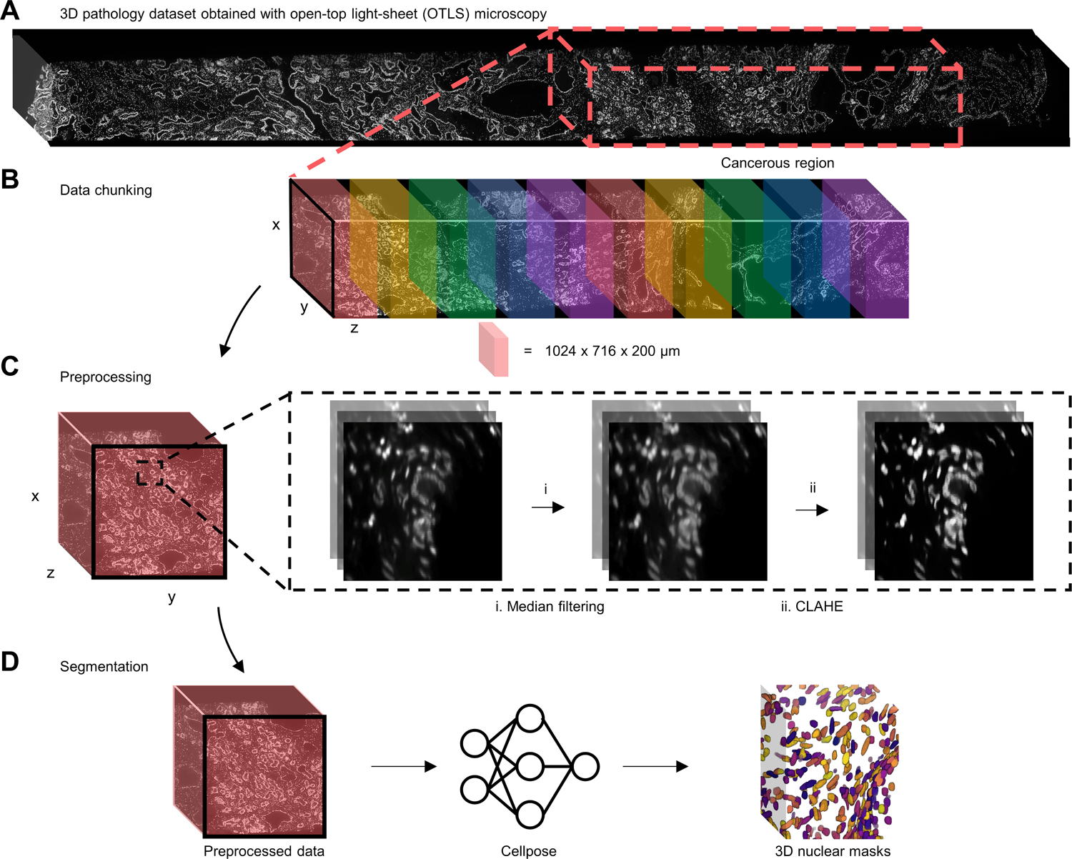
Figure 2. 3D nuclear segmentation pipeline for biopsies imaged with OTLS microscopy.

(A) Nuclear channel (To-Pro-3) of a PCa biopsy imaged by OTLS microscopy with the cancerous region outlined with a dashed red box. (B) The cancerous region is broken up into discrete data blocks before processing. (C) Each data block is passed into a two-step preprocessing procedure before segmentation (see text for details). (D) Preprocessed data blocks are passed into cellpose to generate 3D nuclear segmentation masks, where each segmented nucleus is assigned a unique label.
