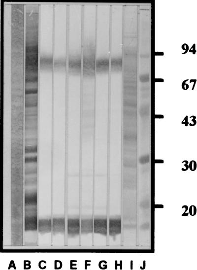
FIG. 1.
Western blot patterns of MAbs from the six specific monoclones against SDS-PAGE-separated V. cholerae O139 strain TH 166 whole-cell Ly. Lanes: A, separated Ly stained by Con A-enzyme conjugate; B through H, Western blot patterns of immune serum of mouse no. 4 and MAbs from clones 12F5-G11, 12F5-G2, 15F5-H5, 5B9-F8, 14C9-D2, and 6C2-D8, respectively; I, separated Ly stained with amido black; J, standard molecular weight markers (numbers at right are relative molecular masses × 10−3).

