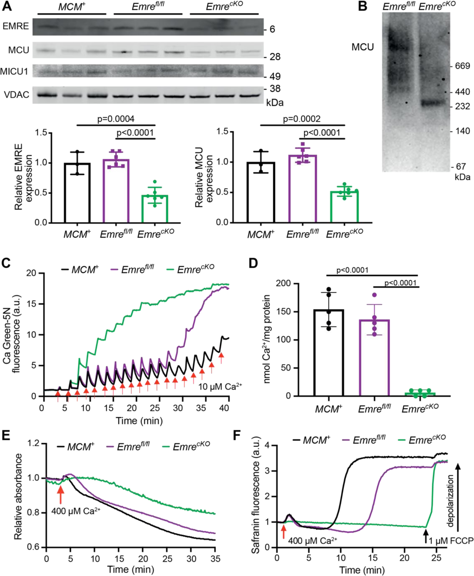
Figure 5. Mitochondrial Ca2+ handling remains impaired with long-term Emre deletion.

Analyses were performed 3 months post-tamoxifen. A) Representative Western blot of EMRE, MCU, and MICU1 in cardiac mitochondria from MCM+, Emrefl/fl and EmrecKO mice. VDAC was used as a loading control. Below, Western blot quantification of mitochondria from MCM+ (n = 3), Emrefl/fl (n = 6), and EmrecKO (n = 6) hearts. Values represent mean ± SD. One-way ANOVA was used for statistical analysis. B) Blue Native gel of freshly isolated mitochondria from Emrefl/fl and EmrecKO hearts, immunoblotted with MCU antibody. Gel is representative of 3 mice (combined males and females) per group. C) Representative Ca2+ retention capacity (CRC) assay in isolated heart mitochondria from MCM+ (black line), Emrefl/fl (purple line) and EmrecKO (green line) mice. The fluorescent Ca2+ indicator Calcium Green-5N was used to monitor extramitochondrial Ca2+. The arrows represent 10 μM Ca2+ added. Traces are representative of n = 5 independent experiments. D) Ca2+ retention capacity calculated from independent traces as shown in (C). The estimated mean Ca2+/mg protein was for MCM+ : 154 ± 30.50, Emrefl/fl : 136 ± 27.01 and EmrecKO : 6 ± 5.47 nmol of Ca2+ / mg protein. Values represent mean ± SD.**p<0.0001, n = 5 in each group. One-way ANOVA test was used for statistical analysis. E) Representative mitochondrial swelling assay monitoring absorbance of isolated heart mitochondria from MCM+ (black line), Emrefl/fl (purple line) and EmrecKO (green line) mice after addition of 400 μM Ca2+. Traces are representative of n = 5 independent experiments. F) Representative traces of membrane potential (△Ψ) depolarization in isolated heart mitochondria from MCM+ (black line), Emrefl/fl (purple line) and EmrecKO (green line) mice after the addition of 400 μM Ca2+. As a positive control in EmrecKO mitochondria, depolarization was induced by addition of 1 μM FCCP. Traces are representative of n = 4 independent experiments. The mean time to Ca2+-induced depolarization, calculated as the time from Ca2+ addition to 50% of the rise in fluorescence, was for MCM+: 10.5 ± 2 minutes, for Emrefl/fl: 12.7 ± 2.3 minutes, and could not be calculated for EmrecKO as FCCP was required for depolarization.
