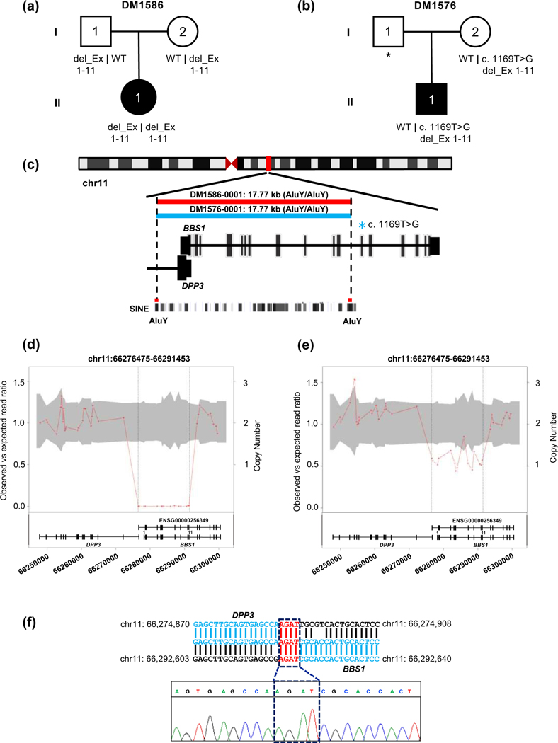
Figure 2. Characterization of a 17.77 kb CNV deletion in BBS1 in family DM1586 and family DM1576.
(a-b) Pedigrees and segregation of BBS1 exon disruptive deletion. (c) Schematic representation of human chromosome 11 and location of BBS1 CNV deletion is indicated with vertical red bar; enlarged view shows schematic of BBS1 transcript and location of AluY-AluY repeats elements. Short interspersed nuclear elements (SINE) (d-e) CNV plot showing homozygous and heterozygous BBS1 deletion, the gray area marks 95% confidence interval and the vertical black dotted lines indicate the location of the CNV; bottom, schematic of BBS1 locus: vertical bars, exons; horizontal line, intronic region; coordinates on chromosome 11 (hg19) are shown. (f) BBS1 breakpoint junction and sequence chromatograms amplified from genomic DNA of DM1586-0001 (II-1); a 4 bp microhomology region is present at the junction of DPP3 and BBS1, highlighted in red.

