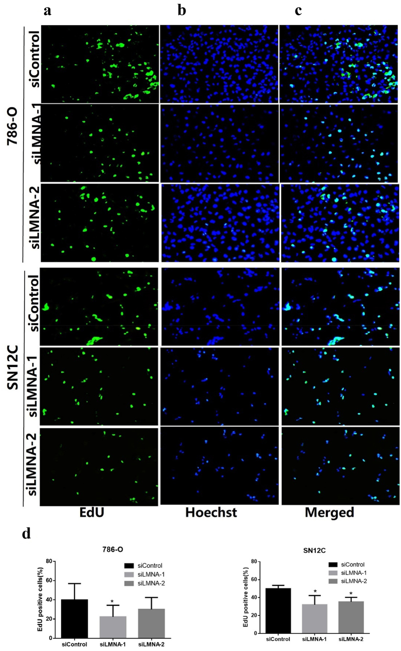
Figure 4.

Representative results of EdU assay after knockdown of LMNA. a: EdU detection showing EdU-positive cells (green) in 786-O and SN12C cells; b: nuclei were stained with Hoechst 33,342 (blue); c: merged images showing EdU-positive cells (Cyan); d) the EdU-positive cells were significantly reduced after knockdown of LMNA in 786-O cells (especially with siLMNA-1) and in SN12C cells. (*p < .05).
