
Figure 1:
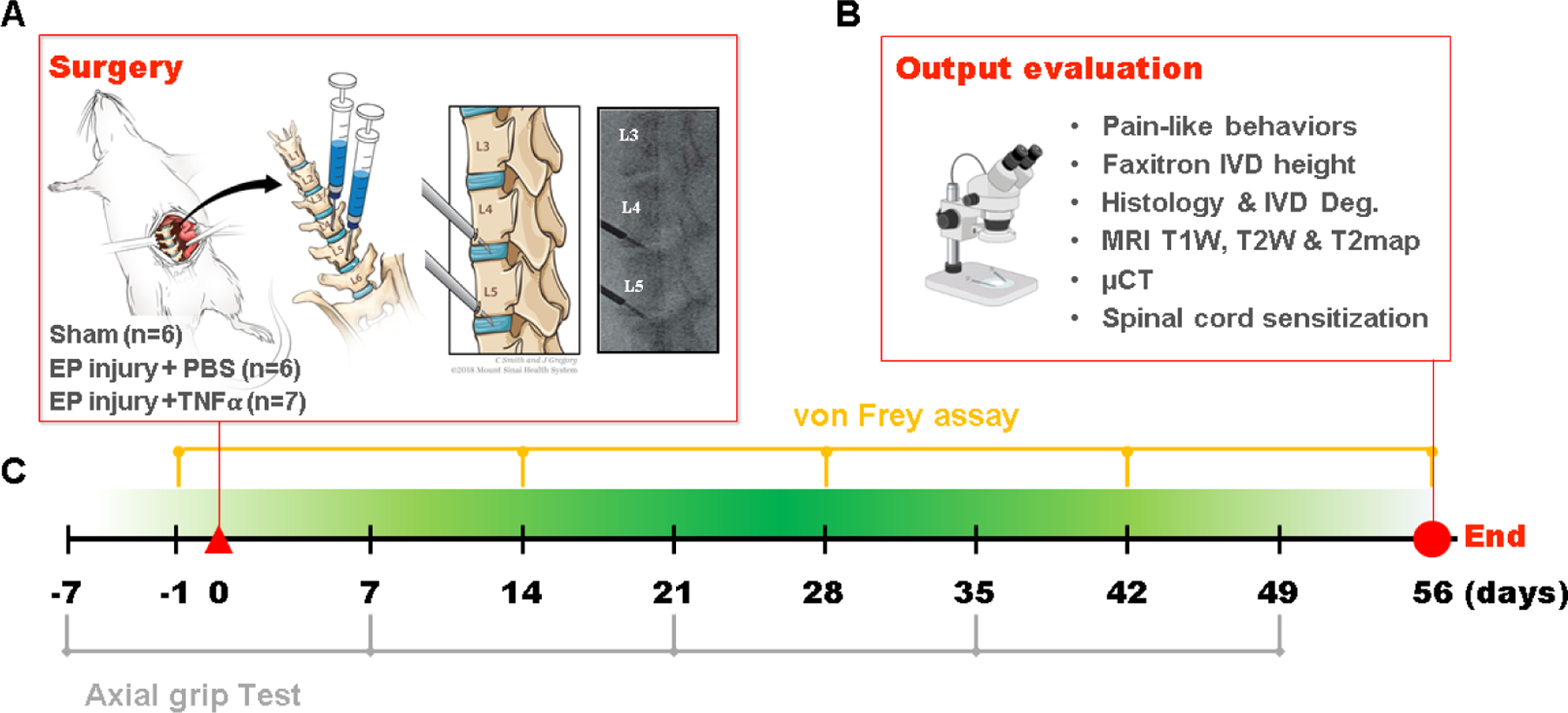
EP microfracture in-vivo model and study design. A) Schematic of procedure with anterior approach. Experimental groups included Sham (n=6); EP injury + PBS injection (n=6) and EP injury + TNFα injection (n=7). Syringes were inserted through a tight-fitting K-wire channel with syringe depth and bevel position confirmed intraoperatively with a C-Arm to be in the center of NP region to enable injectates to be absorbed by the IVD and marrow and minimize the chance of injectate leakage. B) Output variables after t=56 days (8 weeks). C) Timeline of behavioral measurements.