
Figure 4:
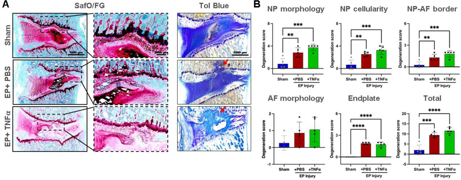
EP caused IVD degeneration. A) Histology with thin sections stained with Safranin-O/Fast green and thick ground and polished sections stained with Toluidine Blue. Red arrows indicating EP defect. B) IVD degeneration grading using Scoring System [49]. **, *** and **** indicate significant differences with p<0.01, p<0.001 and p<0.0001 respectively.