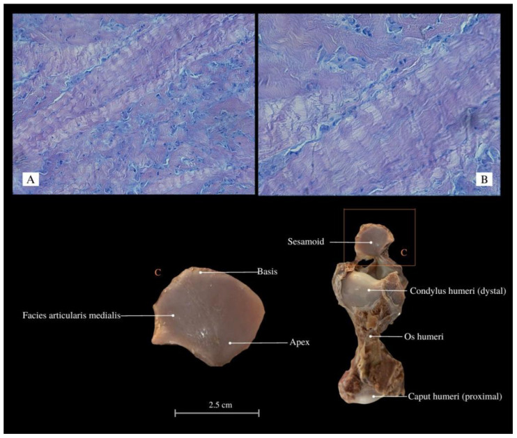
Figure 2.
Histological images (HE stained) of the sesamoid (elastic cartilage, “ulnar patella”) with chondrocytes and chondroblasts arranged in bands between collagen fibers, Mag 200× (A); Mag 400× (B). Macroanatomical views of the sesamoid (C). Apex—top; Basis—base; Facies articularis medialis—medial articular facet; Condylus humeri—condyle of humerus; Os humeri—humerus bone; Caput humeri—head of humerus.

