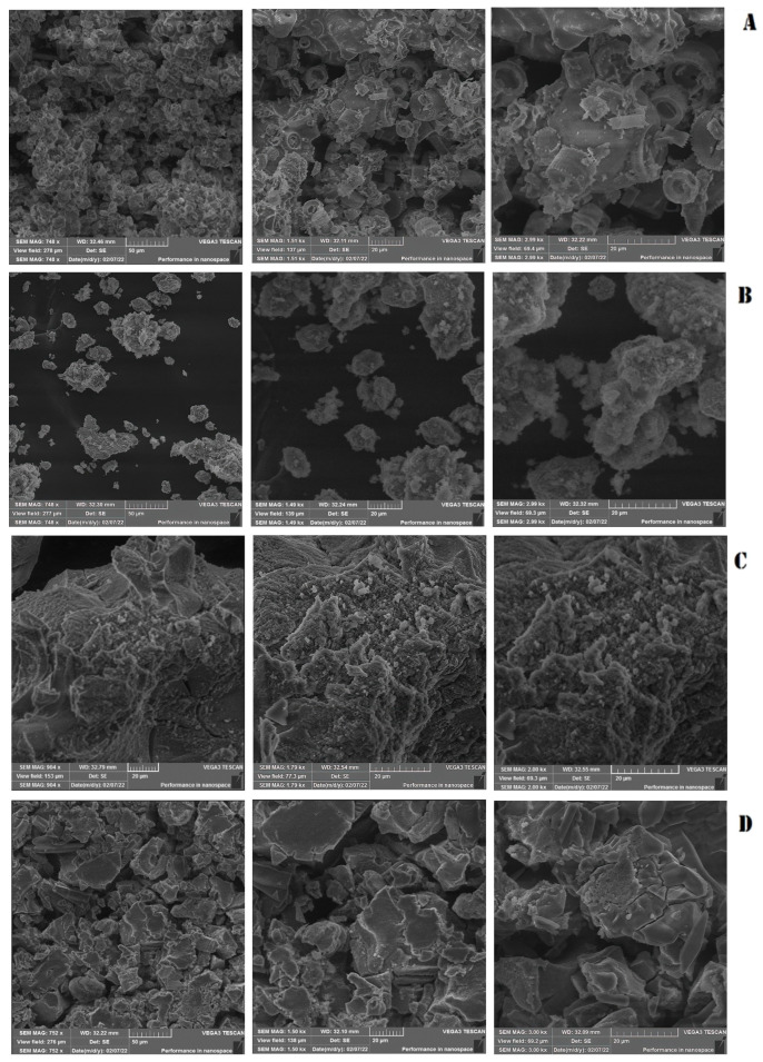
Figure 1.
Scanning electron microscopy (SEM) of silica nanoparticles. (A) Pure silica without treatment. (B) Silica nanoparticles treated with CuO. (C) Silica nanoparticles treated with NaOH. (D) Silica nanoparticles treated with H3PO4. Moving from left to right, the magnification progressively increases.

