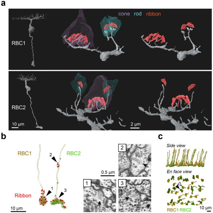
Figure 5. Identification of RBC1 and RBC2 in a SBFSEM volume.
a, Reconstructions of a RBC1 and a RBC2, and zoomed-in images of their dendritic tips at rod and cone terminals. Ribbons in the rod and cones are painted red. b, Ribbon synapse distributions in a RBC1 and a RBC2. The locations of ribbon synapses are marked in red. Arrow heads indicate the locations of example ribbon synapses (arrows) shown in the insets. c, Reconstruction of all RBC1s and RBC2s in the EM volume. Postsynaptic neurons of a centrally located RBC1 (open arrow head) and RBC2 (closed arrow head) were reconstructed in Fig. 6, 7, and S5–9.

