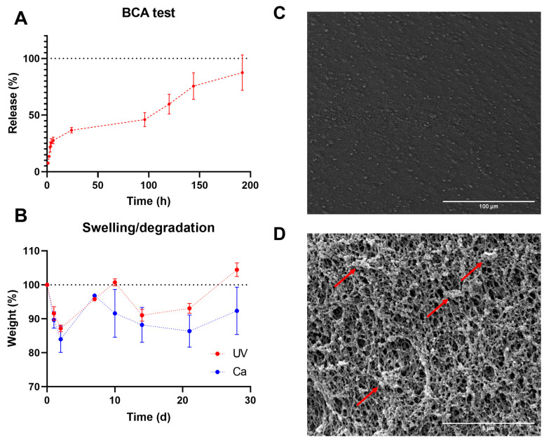
Figure 4.
GA2PL gels’ characterization. (A) BCA test to quantify protein released from UV + Ca-crosslinked GA2PL over 8 days. Red dotted line drawn to guide the eye. (B) Swelling and degradation test of UV- and UV + Ca-crosslinked gels. Blue and red dotted lines drawn to guide the eye. (C,D) SEM images of the surface of GA2PL gels (magnification: 500× and 10,000×, respectively). Red arrows: globular structures attached to the polymer chains. Experimental conditions: GelMA, 5.5% w/v; alginate, 2% w/v; LAP, 0.1% w/v; human platelet lysate, 50% v/v. Data are shown as mean ± s.d., n = 4 replicates.

