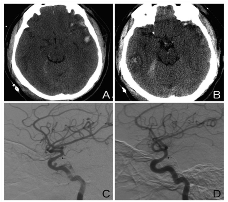
Figure 2.
Illustrative case. A 25-year-old man presented after an assault to the head. An axial non-contrast computed tomography scan of the head showed a left temporal contusion with adjacent subarachnoid hemorrhage (A) and a right temporal contusion and tentorial subdural hematoma (B). Right (C) and left (D) internal carotid artery digital subtraction angiograms (lateral view) obtained on day 10 showed moderate vasospasm of both supraclinoid internal carotid arteries (arrows). Note the traumatic pseudoaneurysm of the cavernous right internal carotid artery.

