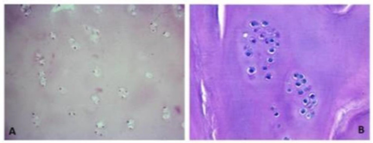
Figure 1.
The histological slides stained with hematoxylin–eosin depict regenerated tissue biopsies obtained two years post membrane-assisted autologous chondrocyte implantation (MACI). The sections reveal limited chondrocytes, which are sparsely distributed either in clusters or in specific regions. These chondrocytes are found embedded within a matrix resembling hyaline cartilage. The images are captured at two different magnifications: (A) 200× magnification and (B) 400× magnification.

