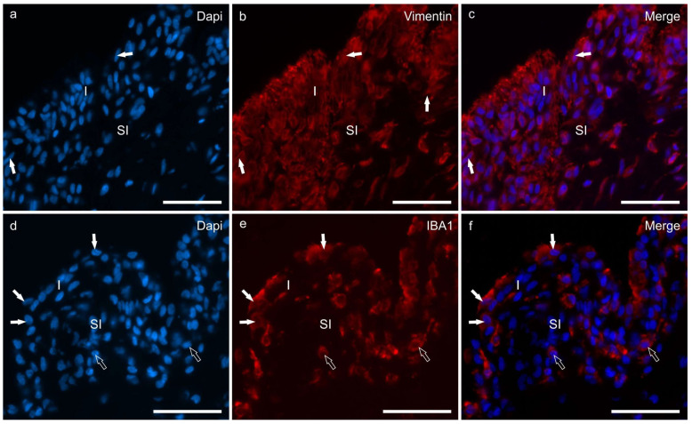
Figure 2.
Photomicrographs of cryosections of the synovial membrane of the stifle joints of dogs showing immunoreactivity for the fibroblast marker vimentin (b) and the macrophage marker IBA1 (e). (a–c) The synovial membrane of the stifle joint showed different layers of synoviocytes (arrows) which expressed moderate-to-bright vimentin immunoreactivity (b). (d–f) Three macrophage-like synoviocytes lining the joint cavity, expressing bright IBA1 immunoreactivity, are indicated by the white arrows (e). The subintimal macrophages (open arrows) also expressed IBA1 immunoreactivity. Abbreviations: I, intima; SI, subintima. Bar: 50 µm.

