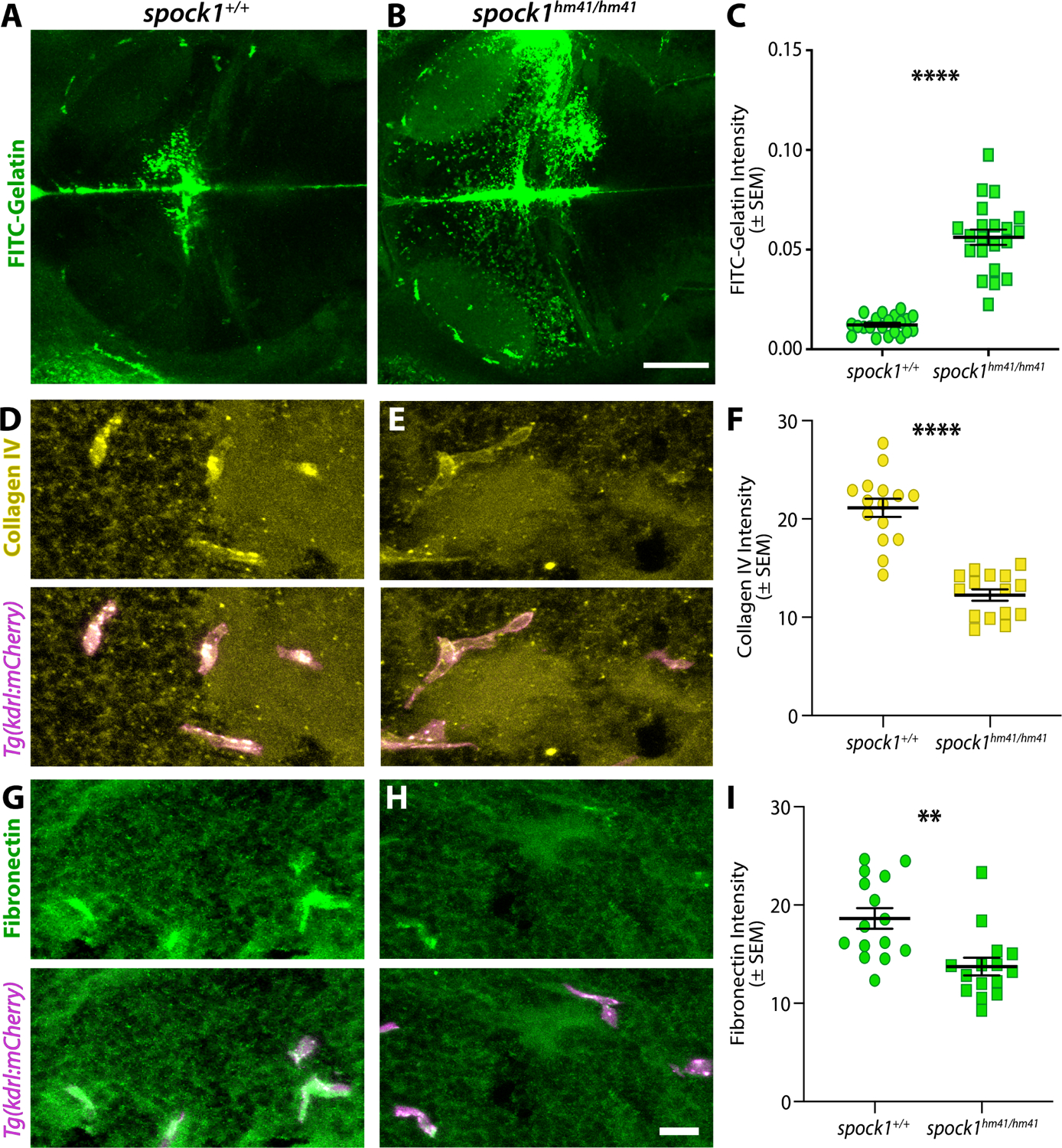
Figure 4. The extracellular matrix is misregulated in spock1 mutants.

(A-C) In vivo gelatin zymography in wild type (A) and spock1hm41/hm41 mutants (B) reveals significantly increased gelatinase activity in the mutant midbrain compared to wild type siblings (C). (D-I) Immunofluorescence staining for vascular extracellular matrix proteins Collagen IV (yellow, D-F) and Fibronectin (green, G-I) that are required for vascular integrity reveal significantly reduced levels of all basement membrane proteins assayed within in the kdrl:mCherry labelled vasculature (magenta), quantified in F and I. N=21 (C) and 15 (F and I) fish analyzed for each genotype and depicted as individual points. Scale bars represent 50 μm (B) and 10 μm (H). ** p=0.0014, **** p<0.0001 by unpaired t test.
