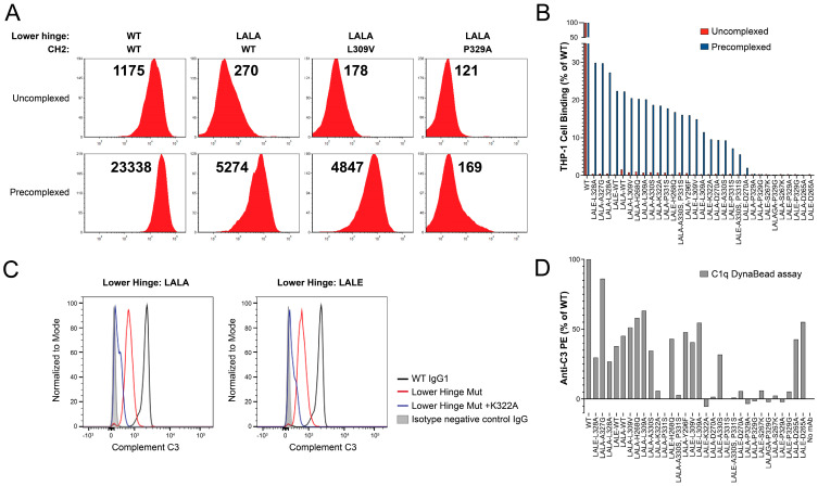
Figure 3.
Elimination of binding to FcγRI and FcγRII presenting THP-1 cells. The THP-1 cells were confirmed to express cell surface FcγRI and FcγRII (Supplementary Figure S1). (A) Representative FACS profiles for WT, LALA, LALA-L309V, and LALA-P329A variants under uncomplexed and pre-complexed conditions. Median fluorescence intensity (MFI) values are shown above each profile. (B) Waterfall bar graph of MFI values collected for the 100 nM ADI-26906 antibody and variants without pre-complexing (red) or with pre-complexing to CD3ε-BSA (blue), conferring multivalent binding, as a percentage of WT. While hinge mutations alone are sufficient to reduce antibody binding under standard assay conditions, only combinations with some, but not all, previously described CH2 mutations are able to eliminate binding under highly avid conditions mediated by pre-complexing to CD3-BSA. (C) Representative C3 binding profiles for the C1q Dyna Bead immune complex assay. Clear separation of C3 activation by WT, lower hinge variants, and lower hinge and K332A combination variants is observed. (D) Bar graph of C3 activation by C1q bound by the antibody immune complex-coated Dyna Beads. The order of antibody variants is the same as in panel (B).

