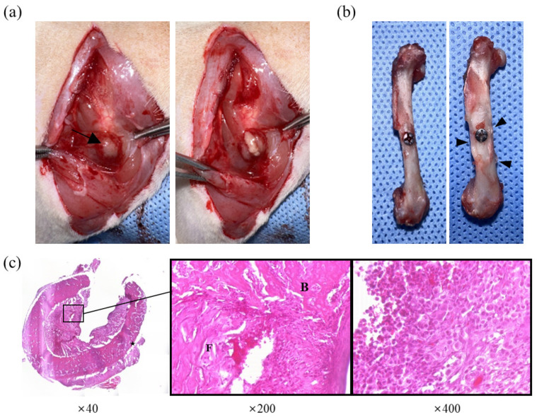
Figure 4.
Observed lesions of group treated with PBS after staphylococcal injection. (a) Gross lesion at the surgical site. Note the abscess formation (arrow) above the femur and purulent exudate erupting from the capsule. (b) Macroscopic cortical changes in femur. Note the thickened cortex around the screw (arrowhead) compared to the femur without macroscopic changes (left). (c) Histological evaluation of osteomyelitis. Enlargement of cortical bone (star) can be observed, and the bone marrow cavity was filled with fibrinous exudate (F), new bone (B), and inflammatory infiltrating cells (right).

