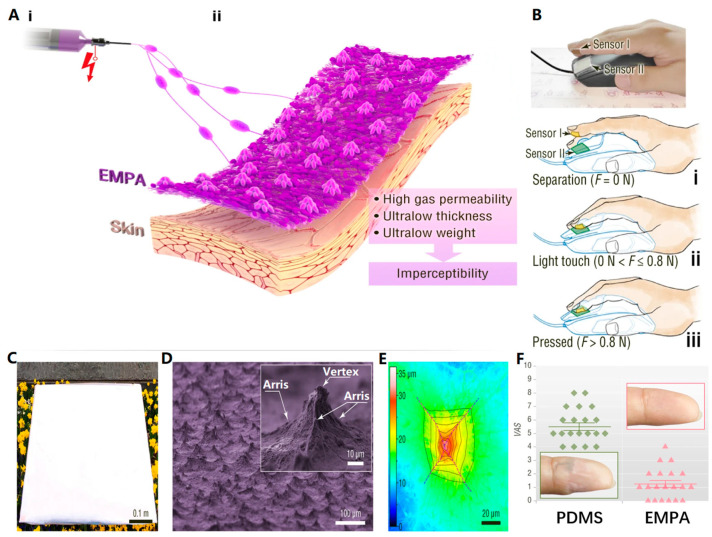
Figure 5.
(A) Schematic illustration of (i) fabrication and (ii) structure. (B) The picture showed the finger operation monitoring of mouse click and three different states: (i) detached; (ii) tapped; and (iii) pressed. (C) Photographs based on large-area EMPA films. (D) SEM image of EMPA; the inset shows a magnified SEM image of an electrospun micropyramid. (E) Laser confocal microscope (LCM) image of electrospun micropyramids; the black dotted line and purple dotted line are the contour lines and alignment lines of the electrospun micropyramid structure, respectively. (F) When both devices were attached to the fingertips, participants reported any sensations, which were assessed on a visual analog score (VAS) (0–10, where 0: no sensory disturbance; 10: extreme discomfort). Horizontal bars represented mean values, and error bars were the standard error of the mean. The insets within the green and pink boxes showed the skin of the fingertip after 7 h of coverage with the different skin devices [43]. Copyright 2022, Springer Nature.

