Figure 5.
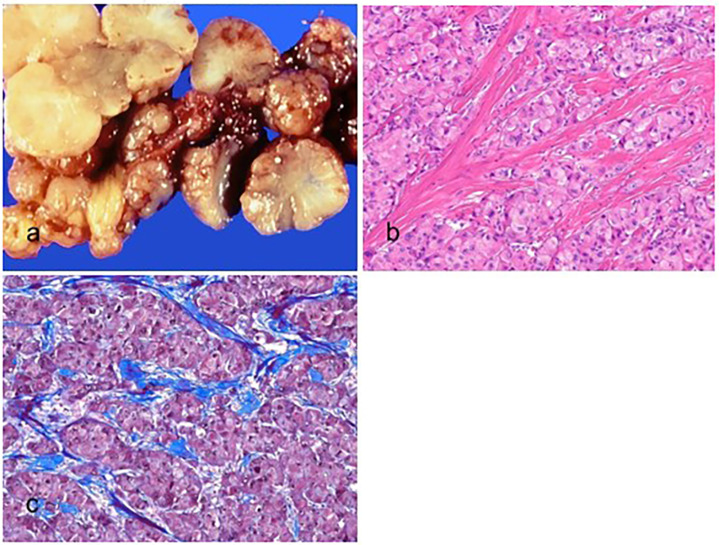
Fibrolamellar carcinoma. (a) The tumor is multilobulated, well-circumscribed, yellow, and firm with a central scarring area. (b) The tumor cells are large, polygonal, and have abundant eosinophilic cytoplasm and prominent nucleoli (hematoxylin–eosin stain, ×100). (c) The intratumoral fibrosis is observed in parallel bands (Masson trichrome stain, ×100).

