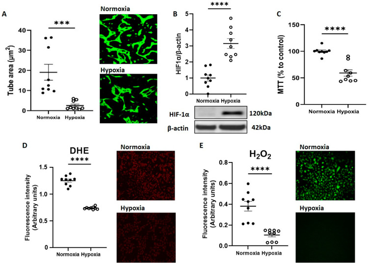
Figure 1.
CB-ECFC angiogenic function and ROS generation were reduced by hypoxia exposure. CB-ECFCs were cultured under hypoxia or normoxia for 48 h prior to endpoint assessment. (A) Matrigel tubulogenesis assay with quantification of the tube area. (B) HIF-1α protein expression determined by Western blot and normalised to β-actin. (C) Cell metabolic activity assessed using MTT assay. (D) Superoxide and (E) hydrogen peroxide production measured by DHE staining and commercially available assay kit, respectively. Representative cell images and Western blots are shown from a single clone for each group. For scatter plots, data were mean ± SEM, n = 9 combined from three CB-ECFC clones; *** p < 0.001, **** p < 0.0001 versus normoxia, unpaired Student’s t-test.

