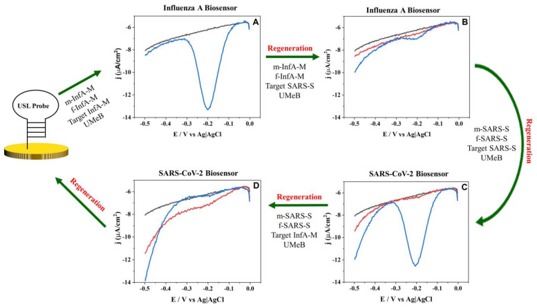
Figure 3.
Regeneration and rehybridization of the USL probe for recognition of InfA-M and SARS-S. Square wave voltammetry response after hybridization with (A) m-InfA-M, f-InfA-M, InfA-M target and UMeB, (B) m-InfA-M, f-InfA-M, SARS-S target and UMeB, (C) m-SARS-S, f-SARS-S, SARS-S target and UMeB, (D) m-SARS-S, f-SARS-S, InfA-M target and UMeB on GDE. Black lines depict the baseline signal; blue lines depict the signal after hybridization or rehybridization of the USL-modified GDE with the indicated strands; red lines depict the signal after washing the 5S-4WJ components from the electrode (regeneration).

