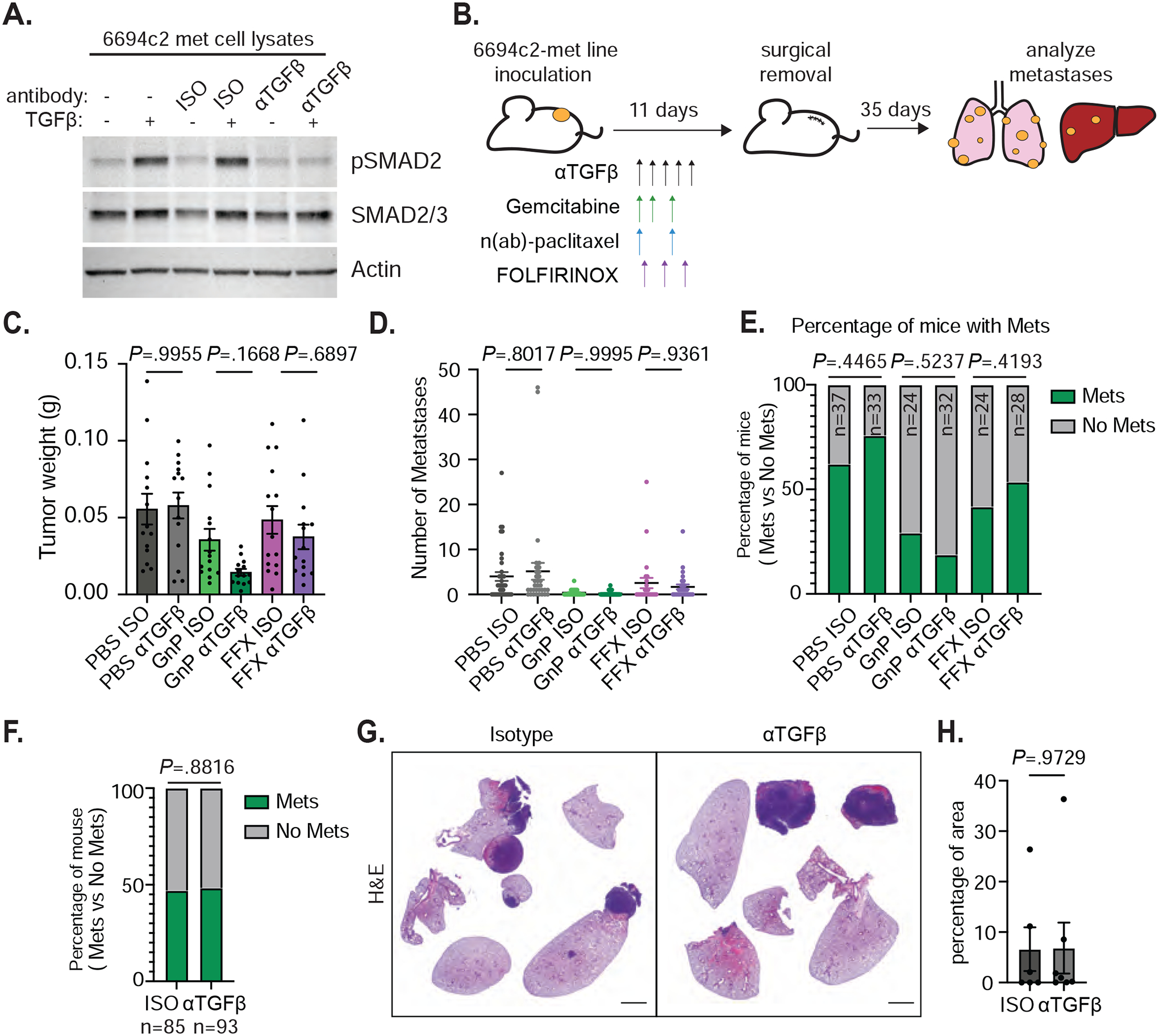
Figure 1: TGFβ blockade does not affect metastasis formation in vivo.

A) 6694c2-met cells were cultured with 5ng/ml TGFβ, and/or 20ug/ml TGFβ blocking or isotype control antibody for 24h. Protein lysates were collected and analyzed by immunoblot. B) Diagram of experimental metastasis model. 180,000 6694c2-met cells were implanted subcutaneously in the lower back of C57BL/6 mice. Mice were treated with αTGFβ or isotype control i.p. every 2 days starting on day 2 for 5 doses. Gemcitabine was given on days 2, 4 and 7; nab-paclitaxel was dosed on days 2 and 7. FOLFIRINOX (5mg/kg oxaliplatin, 50mg/kg irinotecan, 75mg/kg leucovorin, and 75mg/kg 5-FU) was dosed on day 3. Primary tumors grew for 11 days before surgical removal. Metastases in the lung, liver and lymph node were analyzed at day 46. C) Primary tumor weight upon surgical removal. Data are representative of 3 independent experiments. D) Total metastases visually counted in different treatment groups. E) The percentage of mice containing metastases. F) The percentage of mice containing metastases from all isotype-treated or all αTGFβ-treated groups pooled from 3 independent experiments. G) Representative H&E image of metastases in the lungs from isotype and αTGFβ single agent treated mice. H) Quantification was performed on the H&E image for the percentage of area of metastases in the lungs from isotype and αTGFβ single agent treated mice. Scale bars = 2000px. Error bars are SEM throughout. Mann-Whitney t-test was used for comparison between two groups.
