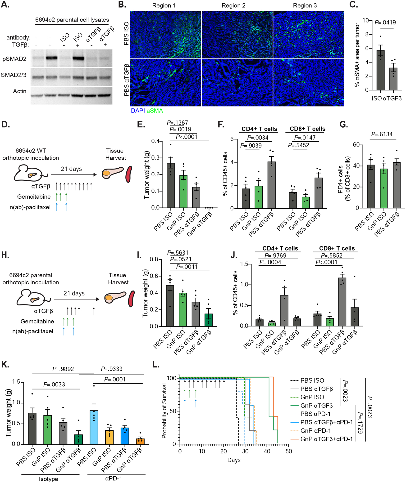
Figure 2: Anti-TGFβ GnP combination treatment is highly effective in poorly immunogenic mouse tumors.

A) 6694c2 parental cells were cultured with 5ng/ml TGFβ, and/or 20ug/ml TGFβ blocking or isotype control antibody for 24h. Protein lysates were collected and analyzed by immunoblot. B) 6694c2 tumors were inoculated into C57BL/6 mice, treated with TGFβ blocking antibody or isotype control and harvested at the midstage of growth on day 15 and frozen in OCT. Representative images from different regions of tumors stained with an antibody against αSMA and DAPI nuclear counterstain are shown. Scale bars = 100μm. C) Quantification of αSMA+ area per tumor. D) Diagram of experimental protocol for orthotopic 6694c2 mouse model for Figure 2E–G. 50,000 6694c2 parental cells were inoculated orthotopically into the pancreas of C57BL/6 mice. E). Tumor weights were measured on day 21. F) Tumor infiltrating CD4 and CD8 T cells were analyzed by flow cytometry of day 21 tumors. G) Percentage of PD1+ tumor infiltrating CD8 T cells. H) Diagram of experimental protocol Figure 2I–J. TGFβ blockade or isotype were dosed i.p. on day 4, day 6, day 8, day 10 and day 20; gemcitabine was given i.p. on day 4, day 7 and day 10; nab-paclitaxel was dosed i.v. on day 4 and day 10. I) Tumor weights were measured on day 21. J) Tumor infiltrating CD4 T cells and CD8 T cells were analyzed by flow cytometry. K-L) 6694c2 tumors were inoculated into mice, treated with TGFβ blockade (every 2 days), PD-1 blockade (twice a week) or corresponding isotype controls until day 20, along with gemcitabine and nab-paclitaxel treatments. K) Tumor weights were measured on day 21; L) Survival of a separate cohort was monitored. N=5 per group. Error bars are SEM throughout.
