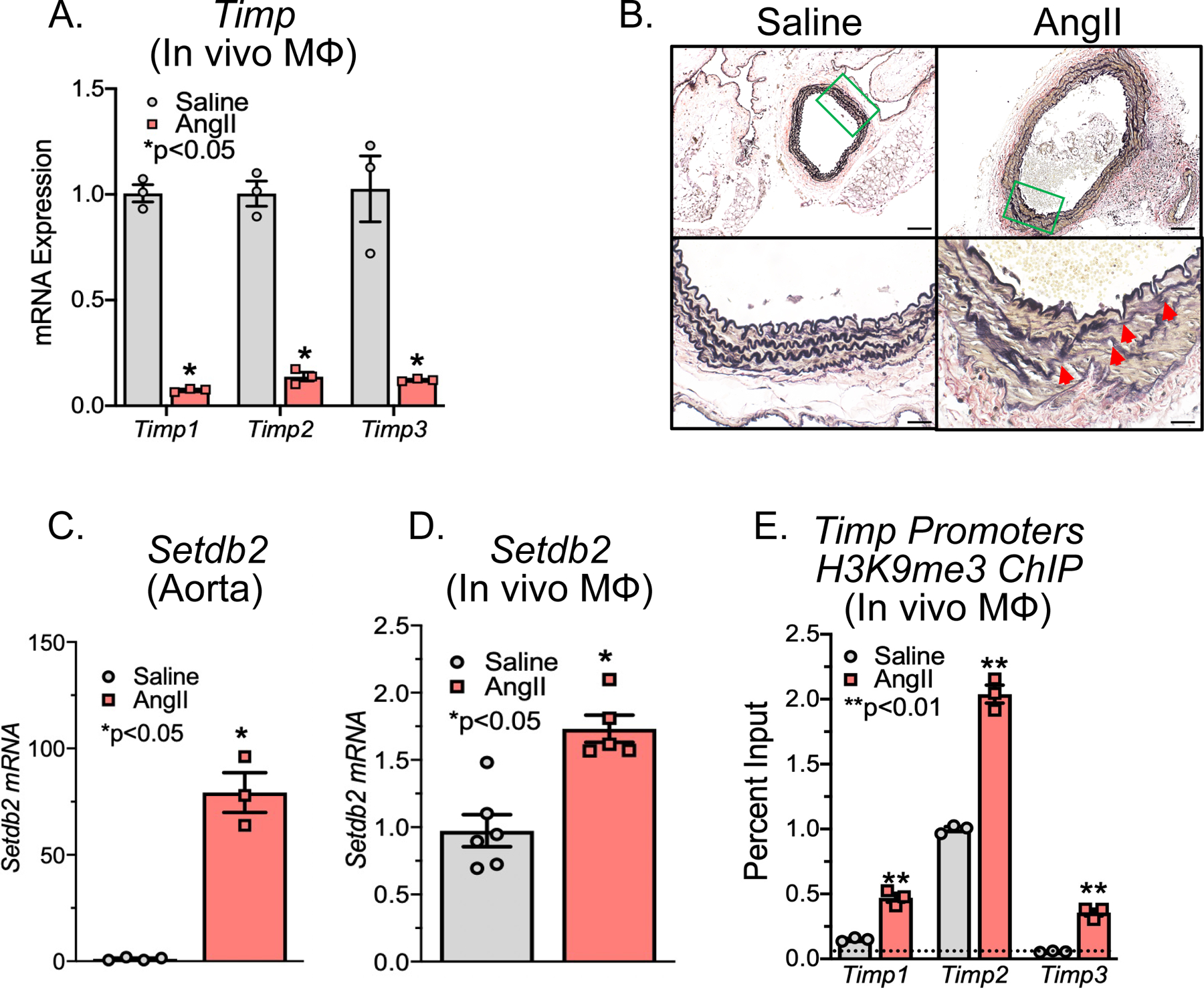
Figure 1. SETDB2 is Increased Murine AAA Macrophages and Increases the Repressive H3K9 Trimethylation on Timp Gene Promoters.

A. Male C57BL/6J mice were injected intraperitoneally with an AAV containing mouse PCSK9D377Y and fed saturated fat diet for 6 wk. Mice were infused with saline or AngII (1,000 ng/min/kg) for 4 weeks. Quantitative PCR analysis of Timp1, Timp2, and Timp3 isolated from in vivo macrophages (MΦ) (CD11b+[CD3−CD19−Nk1.1−Ly6G−]) of mice exposed to saline or AngII for 28 days (n =3/group run in triplicate). *p<0.05, **p<0.01 for Welch’s t-test. B. Representative Verhoeff–van Gieson elastin staining of abdominal aortic sections at 10X and 40X showing disrupted aortic structure in AngII mice compared with saline control mice; scale bar is 50 μm or 10 μm in Verhoeff–van Gieson stain; arrows represent elastin fragmentation. C, D. Quantitative PCR analysis of Setdb2 isolated from aortas or MΦs (CD11b+[CD3−CD19−Nk1.1−Ly6G−]) in mice infused with either saline or Ang II for 28 days (n = 3–4/group run in triplicate). *p<0.05 for Mann-Whitney U test. E. ChIP analysis for H3K9me3 at Timp1, Timp2, and Timp3 promoter was performed (n=3/group run in triplicate). For all ChIP experiments, isotype-matched IgG was run in parallel. Dotted line represents isotype-matched control. **p<0.01 for Mann-Whitney U test.
