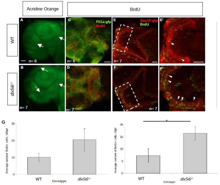
Figure 4.
Proliferation is increased in dlx5i6 mutants at 56 hpf and 5 dpf without changes in cell death. (A,B) Larvae were stained with acridine orange to label apoptotic cells (arrows) at 55 hpf. WT (n = 6, (A)) larvae had few labelled cells as was the case in dlx5i6−/− larvae (n = 7, (B)) in the pharyngeal arches. Mutants had more labelled cells in other parts of the head. (C–F) BrdU staining in 56 hpf (C,D) and 5 dpf (E,F) larvae. Mutants (n = 7 for each timepoint; (D,F)) had overall more BrdU labelled cells compared to WT siblings (n = 6 at 56 hpf, n = 7 at 5 dpf; (C,E)). (E′,F′) 2× zoom of 5 dpf larvae in area within dashed line box. Arrowheads indicate double-labelled cells. (G) Quantification of BrdU+ cells in the MC at 56 hpf (left) and at 5 dpf (right). * p < 0.05, unpaired t-test. Scale = 100 μm.

