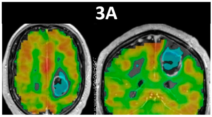
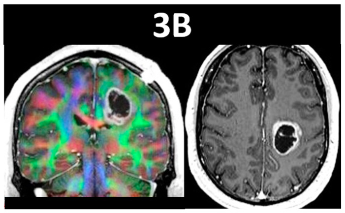
Figure 3.
This figure displays breath-hold cerebrovascular reactivity (BH CVR) maps in the axial and coronal planes in (3A), and DTI color fractional anisotropy (FA) map and anatomic postcontrast T1-weighted image in the coronal and axial planes, respectively, in (3B). This is a 52-year-old male patient with Turcot Syndrome who presented with a WHO grade 3, IDH-wildtype, MGMT-methylated anaplastic astrocytoma that displays prominent NVU, as demonstrated by the Mayo simplified version of the BH CVR task. Notice the regionally reduced CVR superior and lateral to the peripherally-enhancing centrally cystic/necrotic left frontal lobe mass, which resulted in reduced sensorimotor activation. The spatial proximity of the mass to the corticospinal tract (in blue) and the cingulum bundle medially and superior longitudinal fasciculus laterally (in green) is shown in the coronal DTI color FA map. Notice the irregular peripheral enhancement and central cystic and/or necrotic nonenhancing region on the T1-weighted anatomic image.


