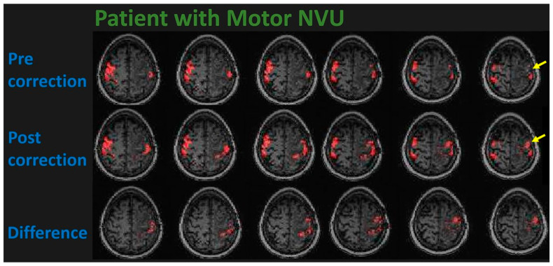
Figure 4.
This figure displays fMRI activation maps in a 58-year-old strongly right-handed male patient with a left frontal lobe WHO grade 4, IDH-wildtype glioblastoma. Although the patient was able to move his right hand well enough to perform the finger tapping and hand opening/closing tasks, virtually no activation is seen in the left precentral gyrus corresponding to the hand representation area of the left primary motor cortex on the initial pre-correction activation maps shown on the (top) row (see yellow arrow pointing to area of expected but markedly reduced activation). The (second) row depicts the post-correction activation map that displays newly visible activation in the primary motor cortex after application of the resting-state amplitude of low frequency fluctuation (ALFF)-based NVU mitigation method. The (third/bottom) row depicts the difference between motor activation on the corrected and uncorrected maps, i.e., the newly-visualized activation resulting from the NVU mitigation method. Please note that the BOLD signal timecourses associated with the newly depicted voxels are identical to those of the originally visible ipsilateral and contralateral activated voxels, indicating high functional specificity of the new activation.

