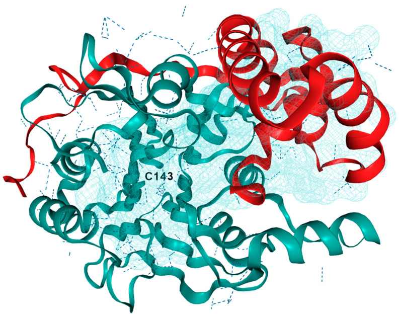
Figure 2.
3D crystal structure of human acid ceramidase (ASAH1, aCDase). PDB ID: 5U7Z. Chain A is red, chain B is teal, and dashed lines indicate hydrogen bonding. The active site is located near Cys143 and a binding pocket near the active site was generated using ezPocket with fconv at 89.4, −3.53, and 203.26 (x, y, z), respectively.

