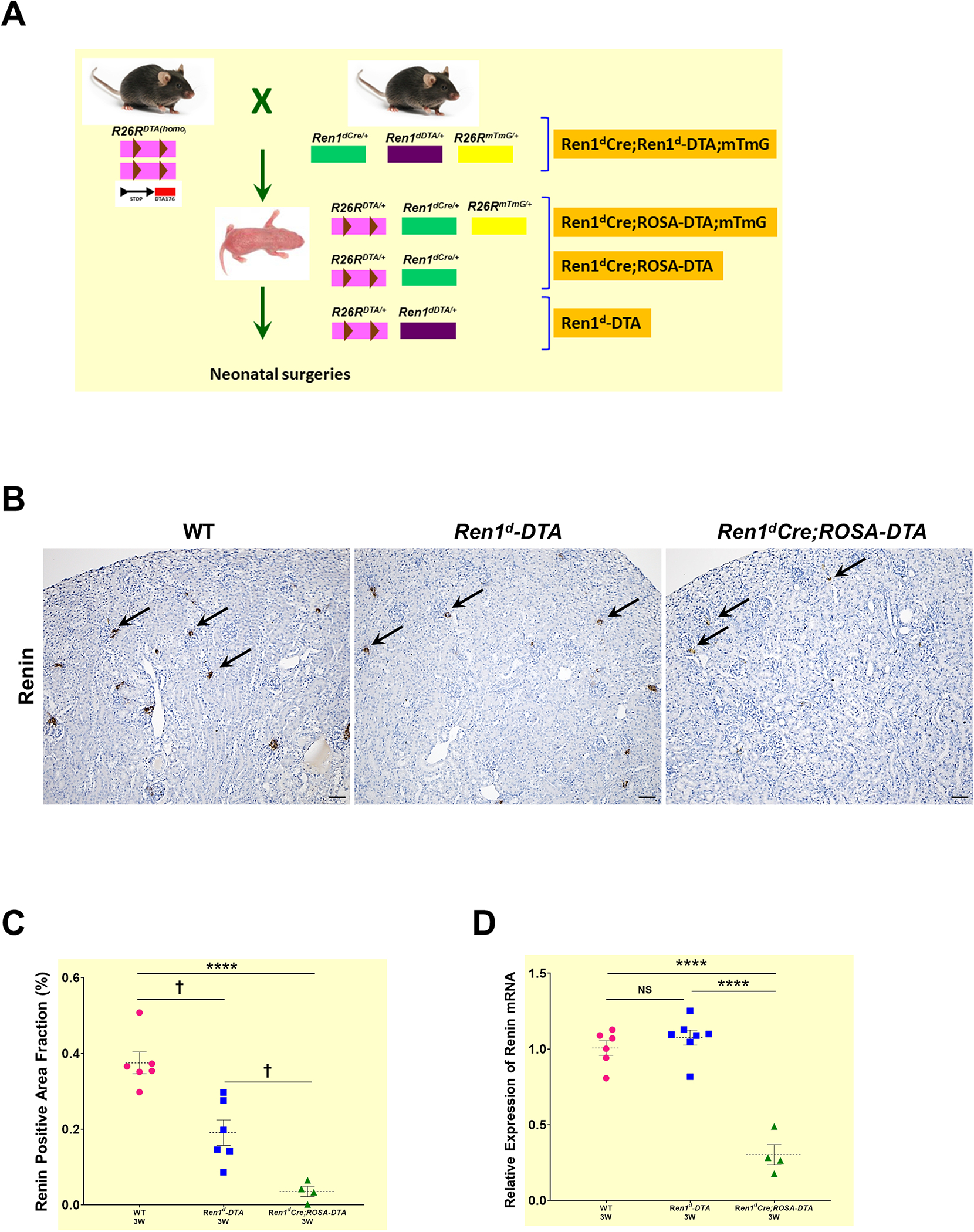
Figure 3. DTA-mediated ablation of renin cells and renin lineage cells:

(A) Crossing strategy to generate neonatal mice expressing DTA in RPCs (Ren1d-DTA) and CoRL (Ren1dCre;ROSA-DTA; Ren1dCre;ROSA-DTA;mTmG) (B) Immunostaining for renin on the kidney sections from 3W old WT, Ren1d-DTA, and Ren1dCre;ROSA-DTA mice not subjected to any surgeries. Renin-positive areas (black arrows) are markedly reduced in the animals expressing DTA compared to the WT group (Scale bar 50μm) (C) Quantification of the renin-positive area fraction in the DTA-genotypes show a reduction which is more pronounced in the Ren1dCre;ROSA-DTA (D) Basal renin mRNA levels analyzed by qRT-PCR showed a significant decrease in the Ren1dCre;ROSA-DTA mice compared to the other two genotypes (*P<0.05; †P<0.01; ****P<0.0001).
