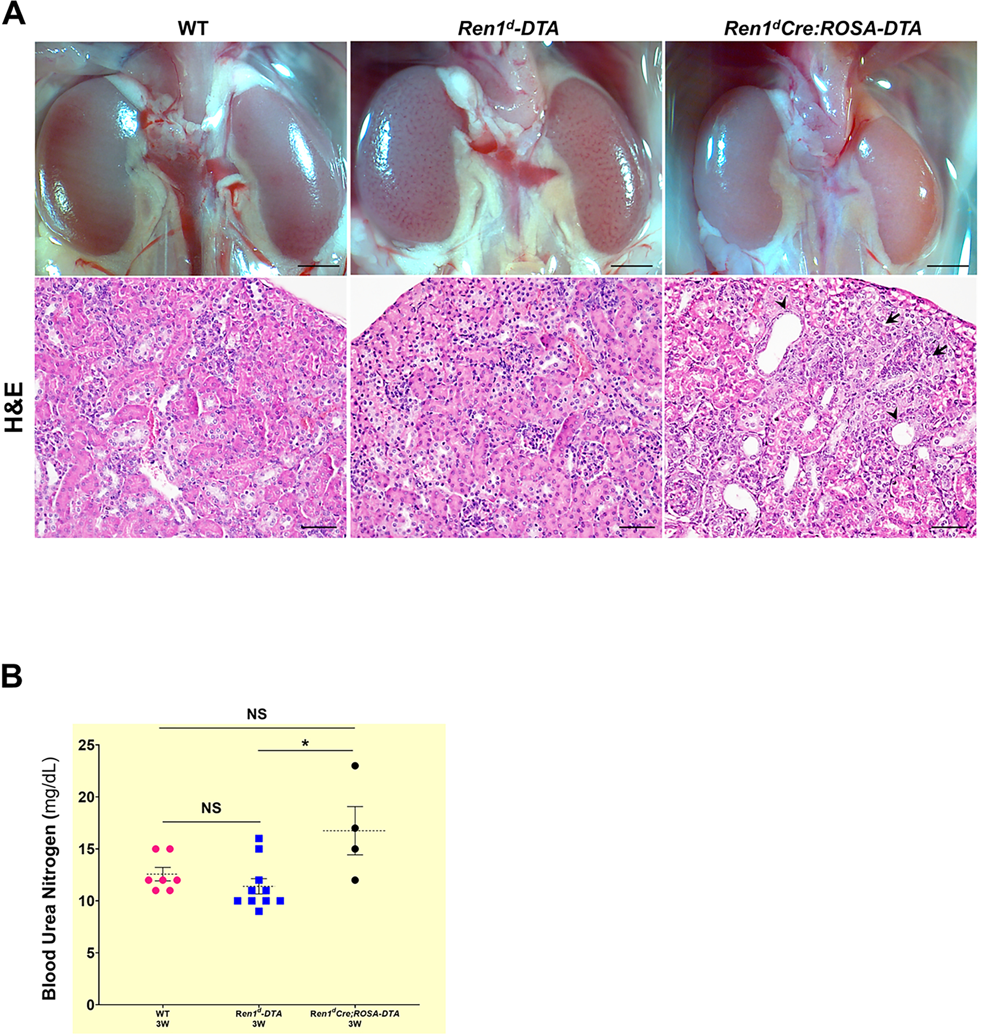
Figure 5. Histological changes due to DTA-mediated ablation of RPCs and CoRL:

(A) Micrographs on whole kidneys and H&E staining on the kidney sections from 3W old WT, Ren1d-DTA, and Ren1dCre;ROSA-DTA animals under basal conditions. Kidneys of Ren1d-DTA mice with intact CoRL showed normal tissue histology similar to the WT group. However, kidney sections of Ren1dCre;ROSA-DTA genotype showed focal sclerotic areas in the renal cortex (black arrows) and tubular dilations (arrowheads) (Scale bar 50μm) (B) BUN levels of the 3W old DTA-positive animals, are not significantly different from the similar age group of WT animals indicating that the kidney function is preserved in the DTA-positive animals (*P<0.05; NS non-significant).
