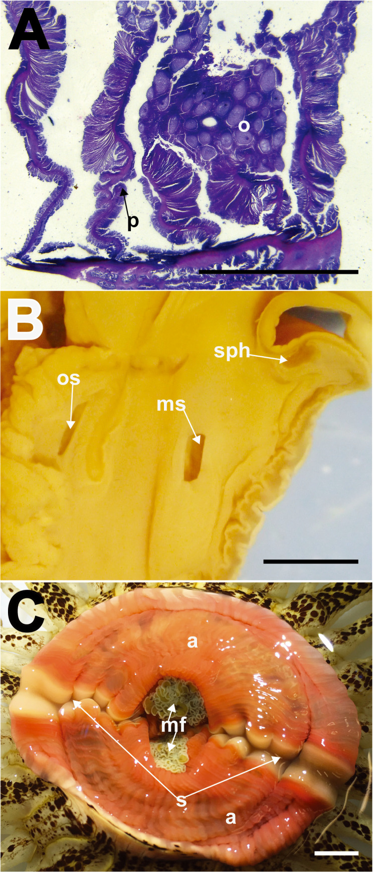
Fig. 5.

Macrodactyla aspera, internal morphology. A, mesenteries of the lectotype (MZC.I.3365), cross section at mid-column. Note the diffuse circumscribed appearance of the retractor muscles, and presence of the retractor pennon. B, transverse section of the distal most end of column (SMNHTAU-Co.7813). Note the presence of a conspicuous, restricted marginal sphincter muscle and the presence of both oral and marginal stomata. C, everted actinopharynx of a live specimen (ZRC.CNI.1090). Note its pinkish appearance, and the presence of a diametric pair of siphonoglyphs. Abbreviations: a, actinopharynx; mf, mesenterial filaments; ms, marginal stomata; o, oocytes; os, oral stomata; p, pennon; s, siphonoglyph; sph, marginal sphincter muscle. Scale bars = 5 mm.
