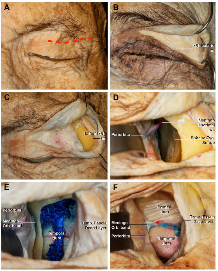
Figure 3.
Macroscopical view of the superior eyelid transorbital endoscopic with the removal of the lateral orbital rim. (A) Skin incision (red dotted line); (B) detail of fibers that form the palpebral portion of the orbicularis muscle; (C) incision of the orbicularis muscle and exposure of the lateral orbital rim. (D) Sub-periorbital dissection. The sphenozygomatic suture and the meningo-lacrimal artery are exposed. (E) After the craniectomy and, in this case, the removal of the lateral orbital rim, the temporal dura and the temporal muscle are exposed; (F) extension of the craniectomy superiorly, with the identification of the frontal dura mater. The meningo-orbital band, which connects the periorbita with the fronto-temporal dura, is shown. Lateral Orb. rim, lateral orbital rim; Meningo Lacrimal art., meningo lacrimal artery; Meningo Orb. band, meningo-orbital Band; Temp. Fascia Deep Layer, temporal fascia deep layer.

