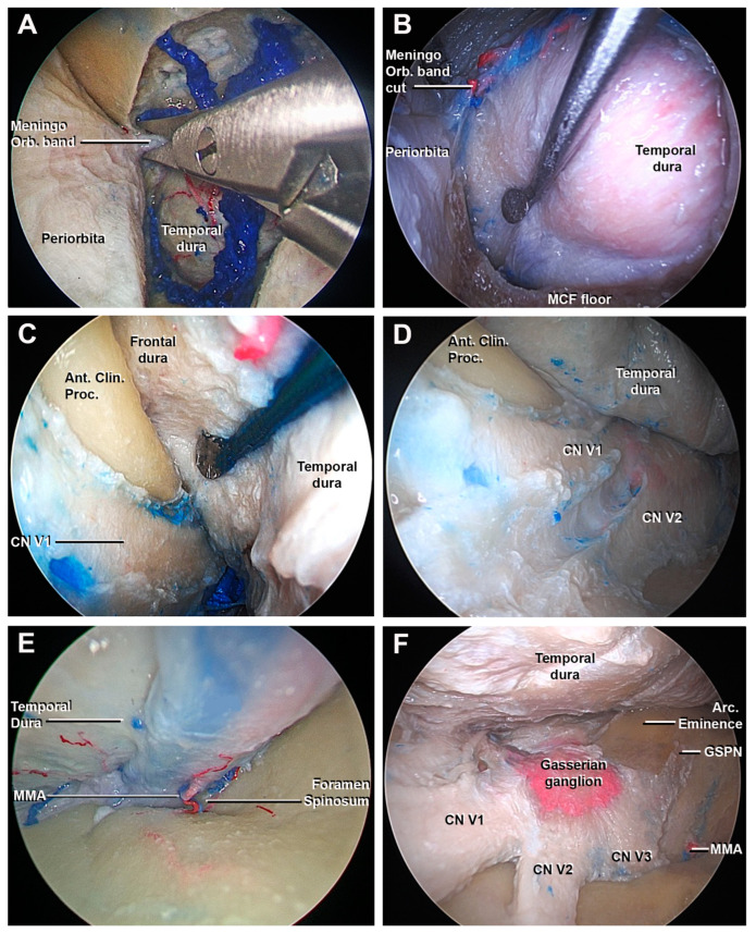
Figure 5.
Endoscopic perspective of the superior eyelid transorbital endoscopic after the craniectomy: endocranial step. (A) The cutting of the meningo-orbital band using a microscissor is performed. (B–D) The interdural dissection between the meningeal layer of the lateral wall of the cavernous sinus and the temporal dura is obtained using a surgical route from lateral to medial: the lateral surface of anterior clinoid process and the cavernous sinus are shown. (E) The middle meningeal artery is found at the level of the foramen spinosum; after the cutting, the extradural peeling of the middle skull base is completed. (F) Final view of the middle cranial fossa: the gasserian ganglion, the three branches (V1, V2, and V3) of the trigeminal nerve, and the GSPN are identified. Ant. Clin. Proc., anterior clinoid process; Arc. Eminence, arcuate eminence; GSPN, greater superficial petrosal nerve; MMA, middle meningeal artery; MCF, middle cranial fossa; Meningo-Orbital band, meningo-orbital band; V1, ophthalmic division of CN V; V2, maxillary division of CN V; V3, mandibular division of CN V.

