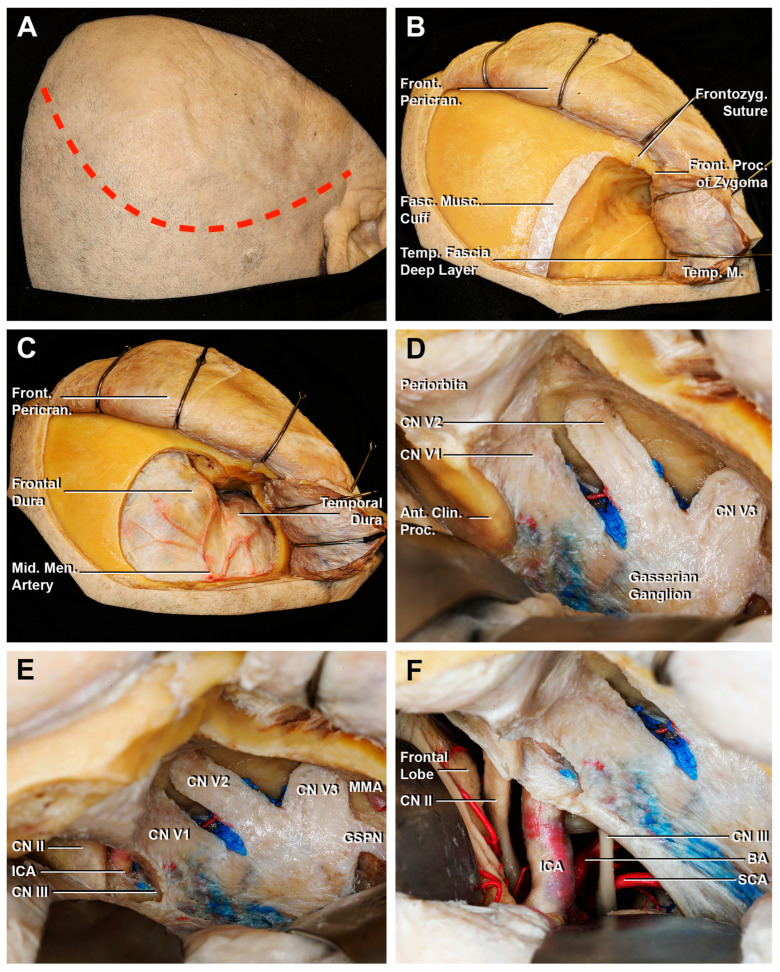
Figure 8.
Key steps of an extradural anterior clinoidectomy through a fronto-temporal craniotomy. (A) A curvilinear skin incision is performed (red dotted line). (B) The temporal muscle is cut, creating the musculo-fascial cuff. Then, the muscle is completely retracted in order to expose the surface for the bone flap. (C) After the fronto-temporal craniotomy, the fronto-temporal dura is exposed. The course of the anterior branch of the middle meningeal artery is shown. (D) The middle fossa dura is elevated until the superior orbital fissure, which exposes the meningo-orbital band. After the cutting of the meningo-orbital band at the level of the sphenoid ridge, the peeling of the dura propria is completed until exposing the bony surface of the anterior clinoid process. (E) After the drilling of the anterior clinoid process, the paraclinoid space is exposed: the clinoid segment of ICA, CN II, and CN III are identified. (F) The opening of the dura allows an intradural surgical view. Ant. Clin. Proc., anterior clinoid process; BA, basilar artery; CN II, optic nerve; CN III, oculomotor nerve; Front. Pericran., frontal pericranium; Front. Proc. of Zygoma, frontal process of the zygoma; Frontozyg. Suture, frontozygomatic suture; Fasc. Musc. Cuff., fascial muscular cuff; ICA, internal carotid artery; GSPN, greater superficial petrosal nerve; Mid. Men. Artery, middle meningeal artery; SCA, superior cerebellar artery; Temp. Fascia Deep Layer, temporal fascia deep layer; Temp. M., temporal muscle; V1, ophthalmic division of CN V; V2, maxillary division of CN V; V3, mandibular division of CN V.

