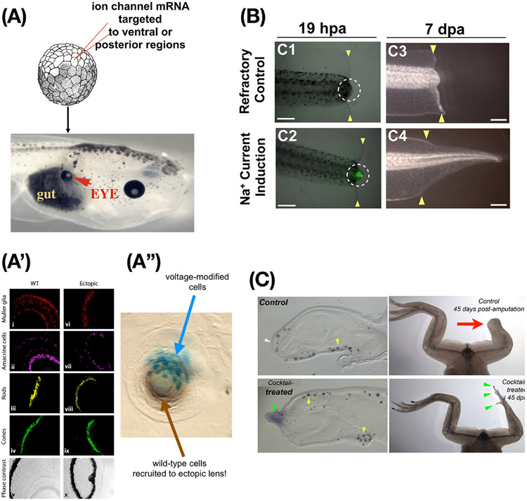
Figure 6: Bioelectric repair strategies.
(A) Misexpressing potassium channels in specific cells to induce the bioelectric prepattern corresponding to a native eye (Fig. 5F) can produce an ectopic eye (red arrowhead) in locations where the “master eye gene” Pax6 cannot do so (posterior to the head); thus, novel differentiation and morphogenesis competencies of cells are revealed by using higher-level (bioelectric) prompts instead of biochemical transcription factor machinery. (A’) Immunohistochemistry confirms all the correct tissue contents of an eye are present despite the very simple nature of the trigger. We did not specify what genes to turn on or how to make an eye; rather, we specified a bioelectric signal that said “make an eye here” and relied on the competency of the cells to do the rest. (A”) When too few cells (labeled in cyan) are injected, the resulting lens includes unmanipulated neighboring cells recruited to help complete the job. (B) Regeneration of the tadpole tail can be induced in non-regenerative conditions by a brief, 1-hour soak in monensin – a sodium ionophore which drives a depolarized wound state (green signal), inducing a tail regrowth program that lasts almost 2 weeks. (C) The same monensin signal induces MSX-1-positive blastema cells and eventually leg regeneration in froglets at a normally non-regenerative stage, showing not only that bioelectric states can induce complex appendage repair in vertebrates, but also that the initial signal does not have to contain much information about the organ to be restored – the host body contains that information, which can be triggered via this interface. Images in A,A’ used with permission from [177]. Image in A”” used with permission from [14]. Images in B used with permission from [90]. Images in C used with permission from [78].

