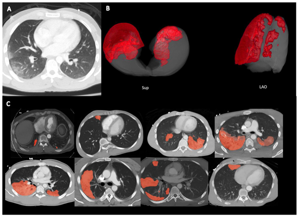
Fig. 1.

A Typical appearance of pulmonary contusion in the right peripheral lung; ground-glass appearance with subpleural sparing. B 3D reconstructed images of pulmonary contusion and lung volume. Sup = superior view; LAO = left anterior oblique view. C Automated segmentation in 8 representative patients. Light red areas are areas of automated segmentation only. Darker red areas are areas of overlap between manual and automated segmentation. Lung volume is denoted with gray outline, corresponding with the pleural margin in all patients illustrated
