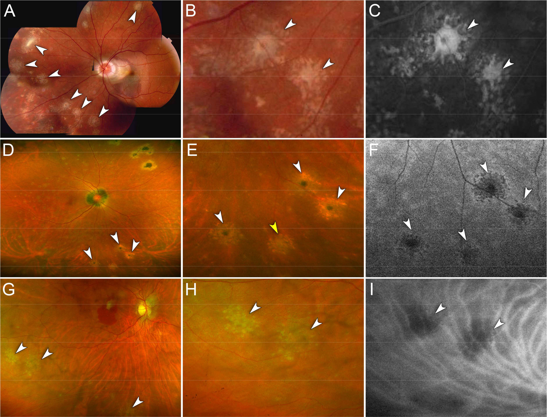
Figure 1. Representative cases of peripheral chrysanthemum lesions on multimodal imaging.

A – C. Case 1. Myopic female in her 30s.
A. Montage color fundus photography of the left eye shows multiple chrysanthemum lesions in the nasal and superior midperipheral retina (white arrowheads). Note the peripapillary subretinal hemorrhage associated with choroidal neovascularization in the macula.
B. Magnified view of (A). Chrysanthemum lesions are distinctively characterized by a grey-yellow central lesion surrounded by satellite dots (white arrowheads).
C. Magnified view of the fluorescein angiogram (late phase) showing hyperfluorescence of the chrysanthemum lesions (white arrowheads).
D – F. Case 11. Myopic female in her 40s.
D. Ultra-widefield pseudocolor fundus photography of the right eye shows multiple chrysanthemum lesions involving the inferior midperipheral retina (white arrowheads). Note the pre-existent punched-out chorioretinal scars in the supero-nasal retina and the peripapillary atrophy.
E. Magnified view of (D). Note the different degrees of pigmentation at the center of the chrysanthemum lesions suggesting the co-existence of recurrent lesions arising from pigmented scars (white arrowheads) and a new lesion with a grey-yellow center (yellow arrowhead).
F. Magnified view of fundus autofluorescence image showing hypoautofluorescent chrysanthemum lesions (white arrowheads).
G – I. Case 12. Myopic female in her 50s.
G. Ultra-widefield pseudocolor fundus photography of the right eye shows multiple chrysanthemum lesions in the inferior midperipheral retina (white arrowheads). Note mild vitritis and a subretinal hemorrhage in the macula associated with choroidal neovascularization.
H. Magnified view of (G). Chrysanthemum lesions are characterized by a grey-yellow central lesion surrounded by pale satellite dots (white arrowheads).
I. Magnified view of the indocyanine green angiogram (mid-phase) showing hypofluorescence of the chrysanthemum lesions (white arrowheads).
