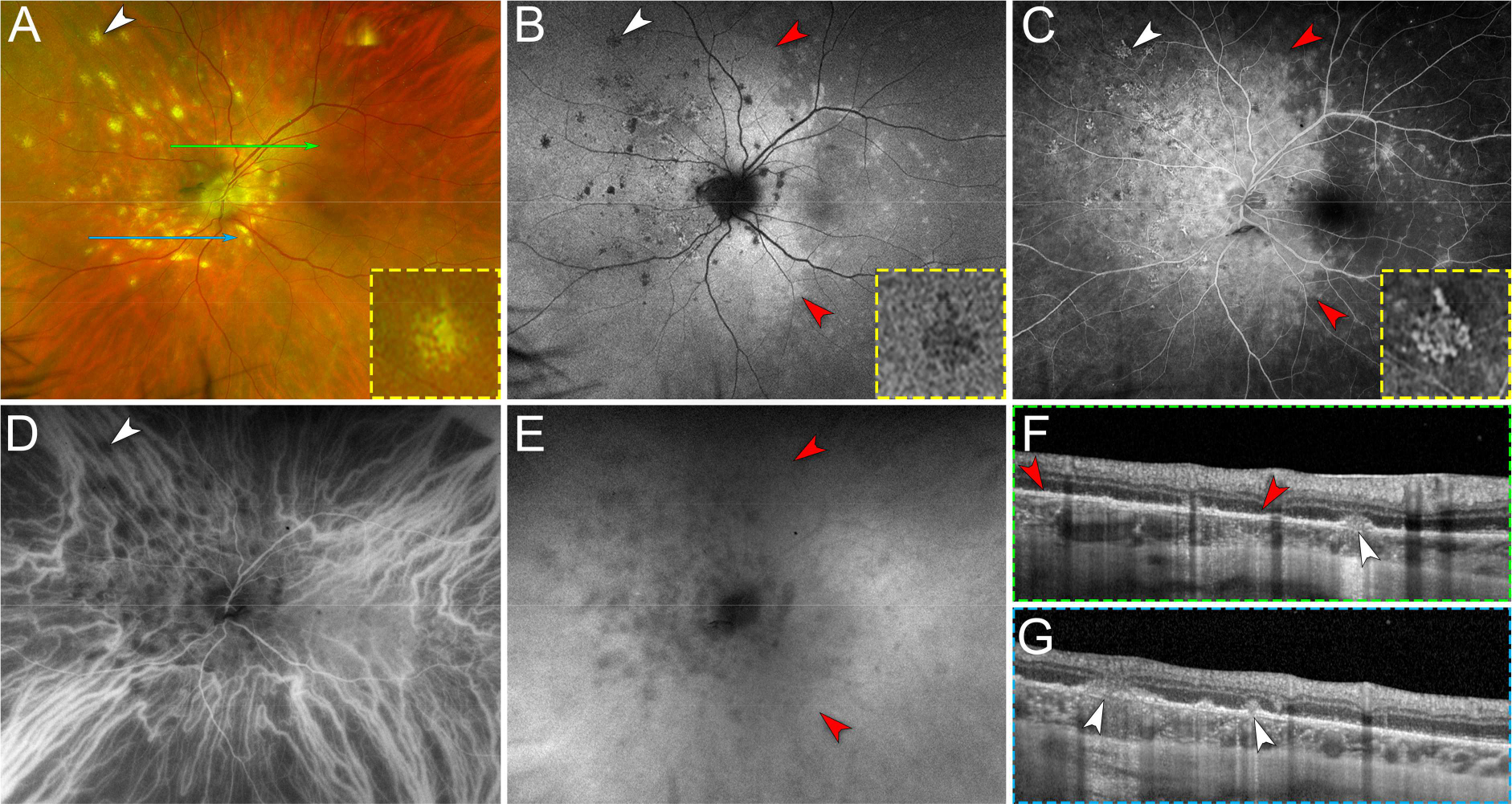
Figure 3. Multimodal imaging of chrysanthemum idiopathic multifocal choroiditis (iMFC) and epiphenomenon multiple evanescent white dot syndrome (EpiMEWDS).

A. Ultra-widefield pseudocolor fundus photography of the left eye shows multiple chrysanthemum lesions in the posterior pole and nasal midperipheral retina (white arrowhead). The inset (yellow dashed box) is a magnified view of the chrysanthemum lesion annotated with the white arrowhead. Note the distinctive morphology of the chrysanthemum lesion characterized by a grey-yellow central lesion surrounded by satellite dots. The green and blue lines indicate the location of the optical coherence tomography (OCT) B-scans displayed in (F) and (G), respectively.
B. Ultra-widefield fundus autofluorescence image shows hypoautofluorescent chrysanthemum lesions (white arrowhead). The inset (yellow dashed box) is a magnified view of the hypoautofluorescent chrysanthemum lesion annotated with the white arrowhead. Note the areas of hyperautofluorescence surrounding the chrysanthemum lesions and the scattered hyperautofluorescent spots corresponding to EpiMEWDS (red arrowheads).
C. Late phase of ultra-widefield fluorescein angiogram shows hyperfluorescent chrysanthemum lesions (white arrowhead) surrounded by confluent hyperfluorescent areas corresponding to EpiMEWDS (red arrowheads). The inset (yellow dashed box) is a magnified view of the hyperfluorescent chrysanthemum lesion annotated with the white arrowhead.
D. Early phase of ultra-widefield indocyanine green angiogram shows hypofluorescent chrysanthemum lesion distributed along and at the tips of the nasal vortex veins (white arrowhead).
E. Late phase of ultra-widefield indocyanine green angiogram shows confluent hypofluorescent areas corresponding to EpiMEWDS (red arrowheads).
F and G. Spectral-domain OCT B-scans show chrysanthemum lesions characterized by subretinal hyperreflective material (white arrowheads) splitting the retinal pigment epithelium/Bruch’s membrane, posterior hypertransmission, focal choroidal thickening and loss of the normal choroidal architecture. Note the areas of ellipsoid and interdigitation zone disruption (between red arrowheads in F) corresponding to EpiMEWDS.
